
Journal of Clinical Images and Medical Case Reports
ISSN 2766-7820
Case Report - Open Access, Volume 2
Unusual inhalation of a tissue piece in an infant
Najoua Imad; Widad Lahmini; Mounir Bourrous*
Department of Pediatric Emergency, Mohamed VI Teaching University Hospital, Cadi Ayyad University, Marrakech, Morocco.
*Corresponding Author: Mounir Bourrous
Cadi Ayyad University, Faculty of medicine and
pharmacy, 7010 Sidi Abbad Street, 40000 Marrakech,
Morocco.
Email: mounirbourrous@yahoo.fr
Received : Nov 19, 2021
Accepted : Jan 06, 2022
Published : Jan 13, 2022
Archived : www.jcimcr.org
Copyright : © Bourrous M (2022).
Abstract
Foreign body inhalation is a major problem in children and is one of the most serious accidents in early childhood. It is considered a common cause of childhood morbidity and mortality.
We present a unique and unusual case of inhalation of a piece of tissue in a 10-month-old infant admitted to the Pediatric Emergency Department of the Mohamed VI Teaching University Hospital in Marrakech in 2021. The patient was initially seen with acute dyspnea, persistent dry cough, and signs of respiratory distress. The auscultation revealed decreased vesicular murmurs in the right lung field. Chest radiography did not reveal the presence of the foreign body, justifying the use of rigid bronchoscopy for diagnostic and therapeutic purposes, and revealing the presence of a tissue in the bronchial area.
Our case report is distinguished by the strange and atypical nature of the foreign body not reported in the literature. Prevention especially parental education is recommended to reduce the morbidity and mortality associated with this domestic accident.
Keywords: foreign body; aspiration; choking; bronchoscopy.
Citation: Imad N, Lahmini W, Bourrous M. Unusual inhalation of a tissue piece in an infant. J Clin Images Med Case Rep. 2022; 3(1): 1562.
Introduction
Foreign Body (FB) inhalation is one of the most critical and common accidents in early childhood due to its high morbidity and mortality in children [1]. FB inhalation has a higher incidence under 3 years of age. Nevertheless, in children under 1 year old, it is the most likely cause of accidental mortalities [1,2]. FB inhalation is often marked by a clinical condition of considerable diagnostic value, which is the foreign body aspiration (coughing, choking and cyanosis) [1,2,3]. It can lead to symptoms and complications that vary according to the nature of the FB, the anatomical site of the enclave and its length of time [2,3]. Therefore, it constitutes a real diagnostic and therapeutic challenge. We report an original case due to the rare nature of the extracted FB.
Case presentation
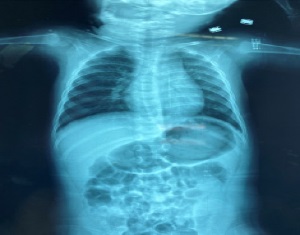
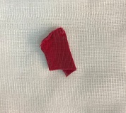
We present the case of a 10-month-old male infant, without any specific pathological history, admitted to the paediatric emergency department of the Mohamed VI Teaching University Hospital of Marrakech with respiratory distress. The investigation with the parents revealed the acute onset of the distress, suggesting a FB aspiration. On clinical examination, the infant was hemodynamically stable, dyspneic with a tachypnea (respiratory rate of 46/min) with the signs of respiratory struggle consisting of supra-sternal pulling and thoraco-abdominal balancing. His room air saturation was 97%. The pulmonary auscultation showed a decrease of breathing movements and vesicular murmurson the right side. The ENT examination revealed a clean oropharynx, with a tongue of normal appearance. The possibility of FB aspiration was considered. Chest X-ray realized did not reveal any radiopaque FB (Figure 1). We performed a rigid bronchoscopy under general anaesthesia which revealed a tissue FB in the right bronchus. Extraction of the piece of tissue was realized without mucosal damage (Figure 2). The patient was administered intravenous amoxicillin-clavulanic acid and nebulized steroids. The evolution was marked by complete resolution of clinical signs after removal of the FB. No complication was observed. The child was discharged to home two days later and was placed on outpatient treatment.
Discussion
The inhaled FB found in chidren are of different types depending largely on cultural and socio-economic factors especially eating habits of countries and patient age. Organic FB, particularly nuts, are the most reported objects; while among inorganic FB, the inhaled FB found in chidren are of various kinds: Toy debris, piece of wood, plastic, magnet, scarf-pin [2- 4]. Of all the inhaled FB types removed in children, tissue is never reported in the literature. The circumstances of foreign body inhalation in children are often accidental. Clinically, the introduction of a FB into the airways is always marked by the penetration syndrome. This, of great semiological value, includes chocking and expulsive coughing [2,5]. Sidell et al. [4] reported that food-object foreign bodies were the most frequent factors associated with choking (94% of all studies). In our patient, the symptomatology was marked by coughing fits and decreased right-sided vesicular murmurs, as well as signs of respiratory struggle.
Imaging may be a diagnostic device for FB inhalation and its complications. The incidence of choice is the frontal chest Xray. It may objectify a radiopaque FB, or may identify indirect signs like notably atelectasis or localised emphysema. In our patient, these signs were absent. In the literature, the chest Xray is normal in variable proportions that can reach 46% [3,5,6]. Note that a normal chest X-ray should never eliminate the possibility of a foreign body. Thus, a very high index of suspicion of FB aspiration should be taken into account in cases with witnessed chocking crisis, acute dyspnoea, wheezing and localized clinical signs. In addition, fluoroscopy can be used as a complement to radiography in children suspected of inhaled FB. It is a sensitive method to show tracheal and bronchial narrowing [7,8]. Computed Tomography (CT) has also been established as an accurate and reliable diagnostic tool in the evaluation of FB inhalation that can increase the rate of positive bronchoscopy [9]. Although the rigid bronchoscopyis generally considered as the gold standard in children with FB aspiration, recent studies have demonstrated the effectiveness and safety of flexible bronchoscopy.
In our case report, we use the rigid bronchoscopy that demonstrated the presence of the FB in the right main bronchus. This was consistent with the literature. Indeed, tracheobronchial FB are mostly lodged in the right main bronchus (72% of all studies) [4]. As to the nature of the FB, we have not found any literature, at the limit of our research, reporting the tissue nature of an inhaled FB in a child, which emphasizes the originality of our case report. The textile fibres cause an inflammatory reaction with exudation by the 24th hour, followed by the formation of granulation tissue (first week), and fibrosis forming by in the second week. This evolution explains, in the absence of infection, the possibility of encystation or even calcifications with a potentially long tolerance [10]. Thus, the rapid extraction must be carried out during the first 24 hours (like in our case) to prevent the occurrence of subsequent complications and avoid damage to the bronchial mucosa. There by, early diagnosis and treatment are of utmost importance to prevent life-threatening and long-term complications. Close communication between the medical team (anesthesiologist, bronchoscopist, pediatrician, and assistants) is essential [11].
Conclusion
Accidental inhalation of FB is a major cause of morbidity and mortality in children. The prevention holds a key place. A national effort and support for preventive education are needed. Education of medical staff and improvement of equipment is also recommended [12,13]. Immediate and adequate multidisciplinary management allows for a better outcome. The originality of our case report lies in the strange nature of the foreign body and its occurrence in an infant at a very early age.
Conflicts of interest: The authors declare no conflict of interest.
References
- Montana A, Salerno M, Feola A, Asmundo A, Nunno ND, Casella F, et al. Risk Management and Recommendations for the Prevention of Fatal Foreign Body Aspiration: Four Cases Aged 1.5 to 3 Years and Mini-Review of the Literature. Int J Environ Res Public Health. 2020; 17: 4700.
- Salih AM, Alfaki M, Alam-Elhuda DM. Airway foreign bodies: A critical review for a common pediatric emergency. World J Emerg Med. 2016; 7: 5-12.
- Ding G, Wu B, Vinturache A, Cai C, Lu M, Gu H. Tracheobronchial foreign body aspiration in children: A retrospective single-center cross-sectional study. Medicine (Baltimore). 2020; 99: e20480.
- Sidell D R, Kim I A, Coker T R, Candice Moreno C, Shapiro N L. Food choking hazards in children. Int J Pediatr Otorhinolaryngol. 2013; 77: 1940-1946.
- Guazzo E, Burns H. Paediatric inhaled airway foreign bodies: An update. Aust J Gen Pract. 2019; 48: 171-174.
- Mathew RP, Liang TIH, Kabeer A, Patel V, Low G, et al. Clinical presentation, diagnosis and management of aerodigestive tract foreign bodies in the paediatric population: Part 2. SA J Radiol. 2021; 25: 2027.
- Ikeda M, Himi K, Yamauchi Y, Ikui A, Shigihara S, Kida A, et al. Use of digital subtraction fluoroscopy to diagnose radiolucent aspirated foreign bodies in infants and children. Int J Pediatr Otorhinolaryngol. 2001; 61: 233-242.
- Huang HJ, Fang HY, Chen HC, Wu CY, Cheng CY, Chang CL, et al. Three-dimensional computed tomography for detection of tracheobronchial foreign body aspiration in children. Pediatr Surg Int J. 2008; 24: 157-160.
- Gibbons AT, Berazaluce AMC, Hanke RE, McNinch NL, Person A, Mehlman T et al. Avoiding unnecessary bronchoscopy in children with suspected foreign body aspiration using computed tomography. J Pediatr Surg. 2020; 55: 176-181.
- Chambi I, Tasker RR, Gentili F, Lougheed WM, Smyth HS, et al. Gauze-induced granuloma (gauzoma): An uncommon complication of gauze reinforcement of berry aneurysms. J Neurosurg. 1990; 72: 163–170.
- Bourrous M, Lahmini W, Nouri H, Haimeur N. Subcutaneous emphysema and pneumomediastinum in child with asthma revealing occult foreign body aspiration: A case report. J Med Case Rep. 2019; 26: 157.
- Ronen O, Kanelo F, Shor D, Ashkar M, Kepten I. Ethnic differences of children with foreign body aspiration: A need for preventive education. Eur Arch Otorhinolaryngol. 2019; 276: 3507-3511.
- Brkic F, Umihanic S, Altumbabic H, Ramas A, Salkic A, Umihanic S, et al. Death as a Consequence of Foreign Body Aspiration in Children. Med Arch. 2018; 72: 220-223.