
Journal of Clinical Images and Medical Case Reports
ISSN 2766-7820
Case Report - Open Access, Volume 2
Single intrauterine foetal death at the second trimester in a twin pregnancy: Case report
Ewane Edwin Nkwelle1*; Djogang Njiomuo Kevin Armel1; Anutebeh Ephesians Nkwetta1; Wateu Pierre2
1 Department of Medicine, University of Buea, Buea, Cameroon, Central Africa.
2 Department of Medicine, University of Bamenda, Bamenda, Cameroon, Central Africa.
*Corresponding Author: Ewane Edwin Nkwelle
Department of Medicine, University of Buea, Buea,
Cameroon, Central Africa.
Email: edwinewane@gmail.com
Received : Oct 27, 2021
Accepted : Jan 07, 2022
Published : Jan 14, 2022
Archived : www.jcimcr.org
Copyright : © Nkwelle EE (2022).
Abstract
A vanishing twin, or foetal resorption, is a foetus in a multi-gestation pregnancy which dies in-utero and is then partially or completely reabsorbed. Single foetal death in the second and third trimester is associated with a higher risk of maternal and foetal morbidity and mortality in the first trimester. The most dreaded maternal complication is Disseminated Intravascular Coagulation (DIC) while the surviving co-twin may be at risk of demise in the same intrauterine environment. This case decribes challenges in management of a second trimester single intrauterine foetal death in a Dichorionic diamniotic (DC) twin pregnancy in a resource-limited setting which resulted in spontaneous vaginal delivery at term with no identified maternal or foetal complications. The management in a limited resource setting is challenging. The risk of prematurity should be weighed against the risk of keeping the surviving co-twin in the same intrauterine environment and management should include psychosocial support.
Keywords: single foetal death; twin pregnancy; case report.
Abbreviations: AC: Abdominal Circumference; BPD: Bi-Parietal Circumference; DC: Dichorionic; DIC: Disseminated Intravascular Coagulation; EFW: Estimated Foetal Weight; EGA: Estimated Gestational Age; FHR: Foetal Heart Rate; FL: Femoral Length; HIV: Human Immunodeficiency Virus; MC: Monochorionic; sIUFD: Single Intrauterine Foetal Death.
Citation: Nkwelle EE, Armel DNK, Nkwetta AE, Pierre W. Single intrauterine foetal death at the second trimester in a twin pregnancy: Case report. J Clin Images Med Case Rep. 2022; 3(1): 1566.
Introduction
It is known that many more twins are formed than born as some are lost during pregnancy, termed “the vanishing twin syndrome” [1]. Single Intrauterine Foetal Death (sIUFD) in twin pregnancy is rare. It presents a management challenge to the obstetricians and is a cause of anxiety for both the pregnant women and the relatives [2]. The prevalence of sIUFD among DC twins is lower than that reported for Monochorionic (MC) twins [1]. The aetiology of sIUFD in twin pregnancies may include twin-to-twin transfusion syndrome, Rhesus incompatibility, congenital abnormalities, preeclampsia, placenta abnormalities, umbilical vein thrombosis, single umbilical artery, umbilical cord knotting, vilamentous insertion, cord entanglement and uterine malformations. In some cases however, the cause may be unknown [3-5].
sIUFD in the second or third trimester poses a dilemma to obstetricians, who may defer intervention as the chance of favourable outcomes exceeds that of poor outcome and intervention may further expose the co-twin to prematurity. The surviving co-twin however needs close follow up to identify any complications [1]. Because of the shared placental circulation in MC twins, the surviving co-twin in MC single foetal demise is at greater risk of complications including exsanguination into the low pressure circulation of the dead twin, shock, anaemia, cerebral injury and possible demise than in DC twins [6]. Passage of tissue thromboplastin from dead foetus and its placenta into maternal circulation can cause maternal Disseminated Intravascular Coagulation (DIC) [7,8].
We present a case of second trimester sIUFD managed conservatively with favourable outcome.
Case presentation
A 23-year-old G3P2002 with a family history of twin pregnancies and no notion of assisted reproduction presented at Malantouen District Hospital for booking visit at 18 weeks of amenorrhea with a large symphysiofundal height of 22 cm and no prior Ultrasound Scan (USS). The physical examination was unremarkable. An obstetric USS was done at 22 weeks of amenorrhea and showed a DC diamniotic twin pregnancy including:
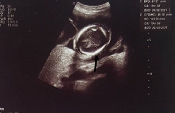
Twin A: the presenting twin, no cardiac activity, Femoral Length (FL): 38 mm; Abdominal Circumference (AC): 138 mm; Biparietal Diameter (BPD): 41 mm; Estimated Foetal Weight (EFW) 350 g, Estimated Gestational Age (EGA): 19 weeks 1 day, placenta anterior (Figure 1).
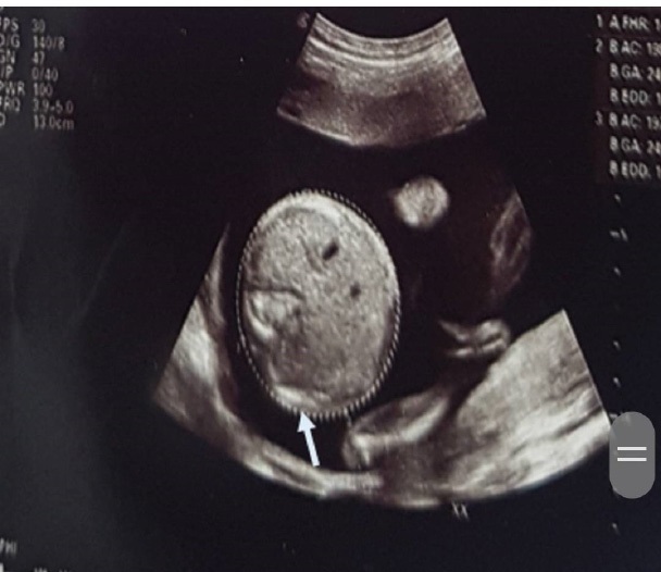
- Twin B: Foetal Heart Rate (FHR): 148 bpm, FL: 42 mm, AC: 198 mm, BPD: 61 mm, EFW: 658 g, EGA: 24 weeks 1 day, placenta posterior (Figure 2).
The laboratory findings were haemoglobin concentration of 10.4 g/L, screening tests for malaria, syphilis, Hepatitis B, Human Immunodeficiency Virus (HIV) and COVID-9 were negative. Her blood group was A Rhesus positive. Follow up for maternal coagulopathy was not done for lack of finances. She was informed of the findings and prognosis.
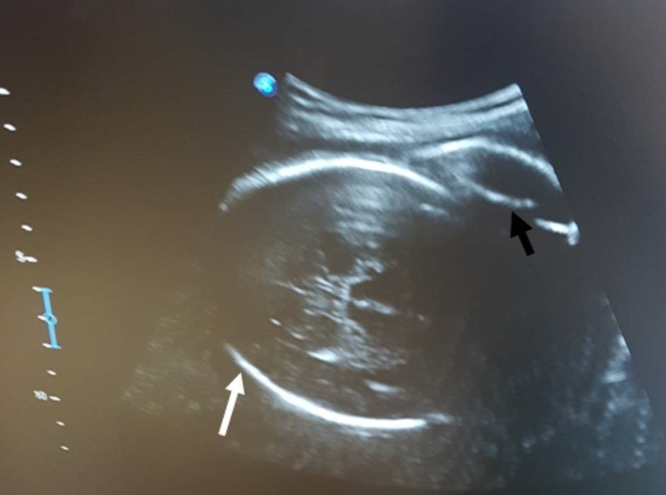
A control USS was done at 34 weeks of amenorrhea which showed a markedly resorbed dead twin in the inferior portion of the uterine cavity and a viable foetus in cephalic presentation with FHR: 137 bpm, EGA: 32 weeks 5 days, EFW: 2102.88 g, placenta posterior (Figure 3). She was trained to monitor daily foetal kick counts for the surviving co-twin. Foetal lung maturation was done using Dexamethasone 6 mg 12 hourly for 48 hours. She was examined twice monthly.
At 37 weeks she presented in the active phase of labour. A control USS showed a viable second twin in cephalic presentation. The first twin was not visualised. The labour led 12 hours 45 minutes later to the delivery of a female baby with first and fifth minute APGAR scores of 6 and 9 respectively, birth weight of 2800g. All other anthropometric measurements were normal. There were no obvious malformations. The cord was 52cm long with no abnormalities. The placenta was regular and weighed 568g. There was no trace of the dead fetus.
Feeding was initiated within the first two hours of life and the patient was discharged two days later.
Discussion
We present a case of sIUFD in a DC pregnancy at about 19 weeks with the dead foetus presenting, which was managed conservatively with a favourable outcome of the co-twin following spontaneous vaginal delivery at term, and the management challenges faced in a resource-limited setting. Because sIUFD in twin pregnancies is rare (between 2.6-5% of twin pregnancies) as is the data on the subject, it often presents a challenge to the caregivers [2,3]. The surviving co-twin may be at risk of the same condition that led to the death of the sibling [9].
Determining the chorionicity in sIUFD is important. The prognosis is more favourable with DC twins [10]. Close follow up is encouraged to identify possible complications on the cotwin and the mother as these are crucial in the management [11]. Circulation of fibrin and thromboplastin from dead foetus in maternal circulation can lead to maternal coagulopathy [8]. Follow up of the mother for coagulopathy was not possible in our setting and delivery in a tertiary centre was not convenient for the patient. She was however counselled on the prognosis. Management of sIUFD should also include steroids to mature the lungs of the co-twin as premature delivery is common. Ward et al showed that sIUFD in DC twin pregnancies with the dead foetus presenting is associated with greater risk of premature delivery [12].
Our plan was to induce labour at term (37 weeks). However, she presented in labour of spontaneous onset at term and the delivery was eutocic. In the absence of complications, it is important to manage conservatively till 37 weeks as earlier interventions may further expose the co-twin to prematurity and its complications [3,7,8]. While MC twins with sIUFD can be more difficult to manage and are delivered mostly between 34 and 37 weeks, management in the DC can be conservative till 37 weeks [1]. The presence of sIUFD alone does not necessitate caesarean section where there is no obstetric indication. In case of obstruction of labour by the presenting dead twin, a caesarean section may however be indicated [2,3,13].
Conclusion
We report a case of sIUFD in a DC twin pregnancy in the second trimester with favourable maternal and neonatal outcome with management challenges in a resource-limited setting. First trimester obstetric ultrasound scan with good resolution is necessary to detect and follow up multiple gestation, and to determine the chorionicity. In sIUFD in DC twin pregnancy, the surviving co-twin can easily be salvaged with close follow up and careful clinical judgement should enable the obstetric caregiver to choose between premature delivery and keeping the co-twin in the same intrauterine environment till term.
Declarations
Competing interests: The authors declare no competing interest.
Authors’ contributions: EN Ewane: Follow up of the patient during antenatal care, labour and delivery, manuscript writing.
NKA Djogang: Follow up of the patient during antenatal care, labour and delivery, manuscript writing.
EN Anutebeh: Manuscript writing and correcting.
P Wateu: Manuscript writing and correcting.
Acknowledgements: We are grateful to Mr Aminkeng Perez for his important role in ultrasound follow up of the patient, and the staff of Malantouen District Hospital for assisting in the close follow up of the patient.
References
- Blickstein I, Perlman S. Single fetal death in twin gestations. Journal of Perinatal Medicine. 2013; 41.
- Haque R, Rahman MM, Akhter K. Intrauterine Single Fetal Demise in Twin Pregnancy. Delta Medical College Journal. 2018; 6: 53–57.
- Yaman Tunç S, Ağaçayak E, Yaman Görük N, İçen MS, Fındık FM , et al. Single intrauterine demise in twin pregnancies: Analysis of 29 cases. Turk J Obstet Gynecol. 2015; 12: 226–229.
- Van den Veyver IBM, Schatteman E, Vanderheyden JS, Van Wiemeersch J, Meulyzer P. Antenatal fetal death in twin pregnancies: A dangerous condition mainly for the surviving co-twin; a report of four cases. European Journal of Obstetrics & Gynecology and Reproductive Biology. 1991; 38: 69–73.
- Gulati N, Mackie FL, Cox P, Marton T, Heazell AEP, Morris RK, et al. Cause of intrauterine and neonatal Death in Twin pregnancies (CoDiT): Development of a novel classification system. BJOG: An International Journal of Obstetrics & Gynaecology. 2020; 127: 1507–1515.
- Klink JMM van, Steenis A van, Steggerda SJ, Genova L, Sueters M, Oepkes D, et al. Single fetal demise in monochorionic pregnancies: Incidence and patterns of cerebral injury. Ultrasound in Obstetrics & Gynecology. 2015; 45: 294–300.
- Akbar DM. Demise of one twin n second trimester with successful outcome of second twin at term. :3.
- Swarankar ML, Shekhawat U, Choudhary M, Choudhary V. Maternal and neonatal outcome of surviving twin after single fetal demise at 25 weeks: A rare case report. Fertility Science and Research. 2017; 4: 5.
- Hillman SC, Morris RK, Kilby MD. Single twin demise: Consequence for survivors. Seminars in Fetal and Neonatal Medicine. 2010; 15: 319–326.
- Arınkan SA, Arısoy R, Api M. Assesment of pregnancy outcomes among twin pregnancies with single fetal demise regarding chorionicity and fetal death time. J Turk Ger Gynecol Assoc. 2019; 20: 147–153.
- Awoyesuku PA, Ohako C, Ngeri B. Conservative management of single fetal death in a twin pregnancy. Int J Reprod Contracept Obstet Gynecol. 2020; 9: 5099.
- Ward PL, Reidy KL, Palma-Dias R, Doyle LW, Umstad MP. Single Intrauterine Death in Twins: The Importance of Fetal Order. Twin Research and Human Genetics. 2018; 21: 556–562.
- Firdouse S, Sanata V, Laxmi K. Fetus papyraceus. IJOGR. 2020; 7: 286–288.