
Journal of Clinical Images and Medical Case Reports
ISSN 2766-7820
Case Report - Open Access, Volume 2
Bony deformities in a child with untreated osteogenesis imperfecta type III
Neeraj Vij; Mohan V Belthur*
University of Arizona College of Medicine, Phoenix, 475 N 5th St., Phoenix, AZ 85004, USA
Phoenix Children’s Hospital, Department of Orthopedics, Main Building, Clinic B, 1919 E, Thomas Rd, Phoenix, AZ 85016, USA.
*Corresponding Author: Mohan V Belthur
Phoenix Children’s Hospital, Department of Orthopedics,
Main Building, Clinic B, 1919 E, Thomas Rd, Phoenix, AZ
85016, USA.
Email: mvbelthur@yahoo.com
Received : Nov 23, 2021
Accepted : Jan 10, 2022
Published : Jan 17, 2022
Archived : www.jcimcr.org
Copyright : © Belthur MV (2022).
Keywords: skeletal dysplasias; radial head dislocation; bone mineralization disorder; fragility fractures; multidisciplinary care.
Citation: Vij N, Belthur MV. Bony deformities in a child with untreated osteogenesis imperfecta type III. J Clin Images Med Case Rep. 2022; 3(1): 1574.
Case description
Osteogenesis Imperfecta (OI) is a skeletal dysplasia that affects the cross-linking of Type 1 collagen leading to a wide spectrum of manifestations including bony abnormalities, scleral abnormalities, dentinogenesis imperfecta, and hearing abnormalities [1].
Orthopedic manifestations of OI include basilar invagination, kyphoscoliosis, codfish vertebrae, bowing deformities of the long bones, coxa vara, protrusion acetabuli, pathologic fractures, and radial head dislocations [1]. The Sillence Classification, originally designed to describe Types I – IV, has been expanded upon to includes Type V, VI, VII, which do not have a Type I collagen mutation, and instead have other genetic explanations (CRTAP, LEPRE1 genes) for the abnormal bone seen on microscopy [1,2]. Type I, IV represent milder forms of disease, Type V-VII represent milder forms of disease, Type III is the most severe survivable form, and Type II is lethal at birth [1,2].
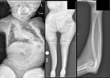
We present a case of Sillence Type III OI, diagnosed by genetic analysis at birth, with significant bony deformities. She did not have appropriate multidisciplinary medical management of her condition and presented to our clinic at age 14. Radiographs of the spine and extremities demonstrated kyphoscoliosis, bilateral protrusio acetabuli, multiapical bowing bony deformities of femora, tibia, humeri, forearm, radial head dislocations, and short stature. This case highlights the importance of multidisciplinary management for children with OI. This includes physical rehabilitation [3], bisphosphonates [3,4] and more recently gene therapies [3,4], and stem cell transplantation therapies [4]. Patients with Osteogenesis Imperfecta Type III have a limited lifespan [5] and it is important to maximize the advantage of medical therapies. In the absence of the appropriate medical care, osteogenesis imperfecta can develop progressive deformities in the long bones, a high risk of fragility fractures, and progressive kyphoscoliosis that can be difficult to manage surgically due to underlying osteopenia.
Final diagnosis: Osteogenesis Imperfecta Type III
Three differential diagnosis:
1. Idiopathic Juvenile Osteoporosis
2. Hypophosphatasia
3. Menkes disease
References
- Ryabykh SO, Popkov DA, Shchurova EN, Ochirova PV, Ryabykh TV. Osteogenesis imperfecta: Current issues of etiology, pathogenesis, classification (systematic review). Genij Ortop. 2021; 27.
- Shaker JL, Albert C, Fritz J, Harris G. Recent developments in osteogenesis imperfecta. F1000 Research. 2015; 4.
- Marini JC, Dang Do AN. Osteogénesis imperfecta - Endotext - NCBI Bookshelf. Osteogenesis Imperfecta. 2020.
- Deguchi M, Tsuji S, Katsura D, Kasahara K, Kimura F, Murakami T. Current overview of osteogenesis imperfecta. Medicina (Lithuania). 2021; 57.
- Paterson CR, Ogston SA, Henry RM. Life expectancy in osteogenesis imperfecta. Br Med J. 1996; 312.