
Journal of Clinical Images and Medical Case Reports
ISSN 2766-7820
Case Report - Open Access, Volume 2
Imaging myocardial perfusion with [99mTc] Tc-HMPAO: Fiction or reality? – Preliminary results
Maria Teresa Faria1*; Maria do Carmo Vilas-Boas2; Paulo Maia2; Pedro Barata3; Ana Oliveira1; Ricardo Rego4; Joel Sousa5; Jorge Pereira1; Francisco Rocha-Gonçalves6; João Paulo Silva Cunha2; Elisabete Martins6,7
1 Nuclear Medicine Department, Centro Hospitalar Universitário de São João, E.P.E., Alameda Professor Hernâni Monteiro, 4200- 319, Porto, Portugal.
2 Institute for Systems Engineering and Computers, Technology and Science (INESC TEC), Faculty of Engineering, University of Porto, Rua Dr. Roberto Frias, 4200-465, Porto, Portugal.
3 Faculty of Health Sciences, University Fernando Pessoa, Rua Carlos da Maia, 296, 4200-150, Porto, Portugal.
4 Neurophysiology Unit, Neurology Department, Centro Hospitalar Universitário de São João, E.P.E., Alameda Professor Hernâni Monteiro, 4200-319, Porto, Portugal.
5 Department of Angiology and Vascular Surgery, Centro Hospitalar Universitário de São João, E.P.E., Alameda Professor Hernâni Monteiro, 4200-319, Porto, Portugal
6 Department of Medicine, Faculty of Medicine, University of Porto, Alameda Professor Hernâni Monteiro, 4200-319, Porto, Portugal.
7 Cardiology Department, Centro Hospitalar Universitário de São João, E.P.E., Alameda Professor Hernâni Monteiro, 4200-319 Porto, Portugal
*Corresponding Author: Maria Teresa Faria
Serviço de Medicina Nuclear do Centro Hospitalar
Universitário de São João, Alameda Professor Hernâni
Monteiro, 4200-319 Porto, Portugal.
Email: mteresafaria@gmail.com
Received : Oct 13, 2021
Accepted : Jan 12, 2022
Published : Jan 19, 2022
Archived : www.jcimcr.org
Copyright : © Faria MT (2022).
Abstract
Introduction and objectives: [ 99mTc] Tc-HMPAO, developed for brain imaging, is taken up by the heart, but never used to study it.
We aimed to compare cardiac images with [99mTc] Tc-HMPAO and [ 99mTc] Tc-Tetrofosmin, using imaging techniques.
Materials and methods: Cardiac gated SPECTs with [99mTc] Tc-HMPAO were compared with myocardial perfusion scintigraphies (MPS) with [99mTc] Tc-Tetrofosmin in three inpatients, from the vascular surgery ward.
We developed algorithms in MATLAB R2016 to compare the [99mTc] Tc-Tetrofosmin/ [99mTc] Tc-HMPAO images. Pixel-wise correlations for slices, reversibility, and polar maps were obtained.
Results: Correlations of both radiotracers’ myocardial images were as high as 0.93. Polar maps correlations were 0.93-0.95 (for both stress and rest) and 0.62-0.90 (reversibility).
One of the patients (smoker) had significant lung [99mTc] Tc-HMPAO uptake.
Conclusions: Cardiac SPECT with [99mTc] Tc-HMPAO might be a screening method for myocardial ischemia in non-smoking patients with epilepsy suspected of having heart changes, and who need to perform a brain perfusion SPECT.
Keywords: myocardial perfusion scintigraphy; epilepsy; HMPAO; myocardial SPECT.
Abbreviations: MPS: Myocardial perfusion scintigraphy; i.v: Intravenous; SA: Short Axis; VLA: Vertical Long Axis; HLA: Horizontal Long Axis; EF: Ejection Fraction.
Citation: Faria MT, Vilas-Boas MDC, Maia P, Barata P, Oliveira A, et al. Imaging myocardial perfusion with [99mTc] Tc-HMPAO: Fiction or reality? – preliminary results. J Clin Images Med Case Rep. 2022; 3(1): 1586.
Introduction
[99mTc] Tc-HMPAO is a radiotracer developed in the 1980s to study regional cerebral blood flow [1-4]. It has been widely used since then, in patients with epilepsy, to localize the epileptogenic zone.
Although this radiotracer’s biodistribution includes the heart [4], to the best of our knowledge, it has never been used to study that organ. The main reason, aside from the excellent existent alternatives, maybe the proximity of the heart to organs with high [99mTc] Tc-HMPAO uptake (lungs and liver) [4,5], potentially interfering with cardiac imaging quality.
Being a brain perfusion radiotracer, mapping of myocardial perfusion seemed also possible, permitting the use of a single radiotracer in patients with epilepsy (known to be prone to cardiac alterations) with less radioactive exposure.
Our aim was to compare myocardial gated SPECT images made with both [99mTc] Tc-HMPAO and [99mTc] Tc-Tetrofosmin, using imaging techniques.
Several authors have compared different radiotracers for myocardial imaging, using varying methodologies [6-12].
We believe this is the first study not only evaluating the possibility of imaging myocardial perfusion with [99mTc] Tc-HMPAO, but also comparing myocardial images with different radiotracers, quantifying the similarities.
Material and methods
We selected 3 inpatients from the Vascular Surgery ward, who already had to perform a myocardial perfusion scintigraphy (MPS) with [99mTc] Tc-Tetrofosmin, as part of their clinical evaluation [one-day protocol, stress/rest, 370 MBq (10 mCi)/1110 MBq (30 mCi), i.v., respectively], and additionally acquired myocardial stress/rest SPECT images with stabilized [99mTc] TcHMPAO [two-day protocol, 555 MBq (15 mCi), i. v., each]. The stress agent was adenosine for both studies and images were acquired in gated mode, 40 min after the i.v. injections.
The interval between each radiotracer scintigraphy varied between 4 months and 1 year, and no major cardiac event was registered in that period, except for patient 3, who developed heart failure.
All studies were processed with a cardiac dedicated software (QGS/QPS), providing three reconstructed planes (short axis – SA –, vertical long axis – VLA –, and horizontal long axis – HLA) and exported into DICOM files.
Due to the inexistence of a heart model on which to calculate normally used clinical parameters, a quantitative analysis of [99mTc] Tc-Tetrofosmin and [99mTc] Tc-HMPAO image similarity is proposed here.
Processing algorithms were developed in MATLAB R2016 to compare the [99mTc] Tc-Tetrofosmin and [99mTc] Tc-HMPAO images of each reconstructed plane (SA, VLA, and HLA).
For image pre-processing, the first and last two slices of the reconstructed images were removed, since no important heart structures were clearly visible. Then, we applied a 2-dimensional median filter, which replaces each pixel by the median value of its 1 by 1 neighbourhood. Due to low image resolution, higher window sizes lead to a significant distortion of the image shape, so we found 1 by 1 value to be optimal for the available dataset.
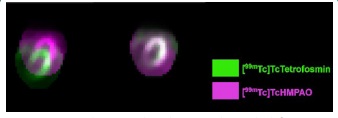
Afterwards, images were aligned, applying the affine transformation that maximized the mutual information [13] between [99mTc] Tc-HMPAO and [99mTc] Tc-Tetrofosmin. Figure 1 exemplifies the initial and final results.
Then, we thresholded the images by selecting all pixels above an intensity of 0 as the region of interest, and identified the bounding box (minimum rectangle on which all image points are included. The purpose was to calculate image correlation metrics corresponding only to this region. We calculated a pixel-wise correlation for every slice, using Pearson’s Correlation Coefficient.
Reversibility of the defects was also evaluated (correlation of the rest-stress subtraction with each radiotracer), as were the polar maps.
We analyzed the LV ejection fraction (EF) and volumes (end diastolic and end systolic) with QGS, in every patient, with both radiotracers.
Ethical approval
This study was approved by our Institution Ethics Committee, as it was in accordance with their ethical standards and with the 1964 Helsinki declaration and its later amendments. All participants gave their informed consent.
Data availability
All datasets generated during and/or analysed during the current study are available from the corresponding author on reasonable request.
Results
We evaluated 3 patients with stress/rest images with both [99mTc] Tc-HMPAO and [99mTc] Tc-Tetrofosmin.
All of them had high correlations for perfusion images (Figure 2), better in the VLA: 0.88 to 0.93 (stress), and 0.88 to 0.90 (rest). Reversibility correlations (Table 1, Figure 3) varied between 0.46 and 0.80 (3 axes).
Gated images correlations between each slice of each gate were also better in VLA [mean values: 0.78-0.89 (stress), 0.78- 0.84 (rest)]. Figure 4 shows the correlations of gated images for all patients.
Table 1: Correlation of reversibility (3 axes) between [99mTc] Tc-HMPAO and [99mTc] Tc-Tetrofosmin images, for patients 1, 2 (smoker) and 3. |
|||
Patient |
Correlation |
||
Short Axis |
Vertical Long Axis |
Horizontal Long Axis |
|
1 |
0.76 (0.08) |
0.80 (0.06) |
0.79 (0.08) |
2 |
0.46 (0.20) |
0.54 (0.07) |
0.47 (0.069) |
3 |
0.63 (0.06) |
0.68 (0.10) |
0.71 (0.12) |
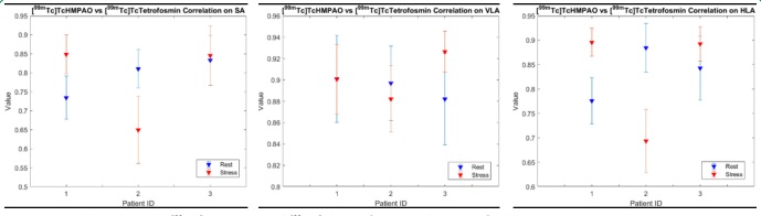
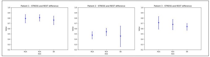
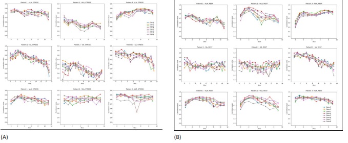
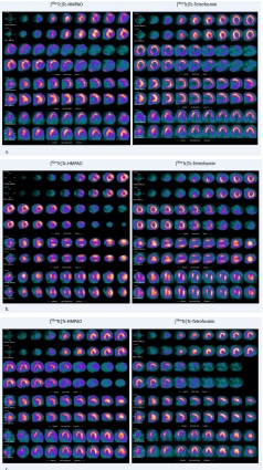
Table 2: Correlation of [99mTc] Tc-HMPAO and [99mTc] Tc-Tetrofosmin polar maps: stress, rest, and reversibility, for patients 1-3 |
|||
|
Polar Maps Correlation |
||
Patient |
Stress/Stress |
Rest/Rest |
Reversibility |
1 |
0.93 |
0.95 |
0.82 |
2 |
0.95 |
0.93 |
0.62 |
3 |
0.94 |
0.94 |
0.90 |
Table 3: Left Ventricle Ejection Fraction and volume values for each patient with [99mTc] Tc-HMPAO and [99mTc] Tc-Tetrofosmin. |
|||||
Subject |
Evaluated Parameter |
Stress |
Rest |
||
[99mTc]Tc-HMPAO |
[99mTc]Tc-Tetrofosmin |
[99mTc]Tc-HMPAO |
[99mTc]Tc-Tetrofosmin |
||
P1 |
EF (%) |
23 |
20 |
20 |
23 |
ESV (mL) |
113 |
183 |
124 |
159 |
|
EDV (mL) |
147 |
230 |
156 |
207 |
|
P2 |
EF (%) |
51 |
51 |
40 |
50 |
ESV (mL) |
32 |
55 |
55 |
56 |
|
EDV (mL) |
65 |
112 |
91 |
111 |
|
P3 |
EF (%) |
53 |
27 |
55 |
30 |
ESV (mL) |
35 |
62 |
47 |
67 |
|
EDV (mL) |
74 |
84 |
105 |
96 |
|
The LV ejection fraction (EF) with both radiotracers was equivalent in almost all patients (Table 3). The exception is Patient 3, whose EF calculated with [99mTc] Tc-Tetrofosmin was significantly lower (severely compromised) when compared to the one calculated with [99mTc] Tc-HMPAO (normal). Patient 2 had only one value significantly different from the others, which was the rest EF evaluated with [99mTc] Tc-HMPAO (mildly compromised compared to normal).
LV volumes with [99mTc] Tc-HMPAO were generally inferior to the ones calculated with [99mTc] Tc-Tetrofosmin.
Discussion
[99mTc] Tc-HMPAO mechanism of localization in the brain is related to its lipophilic nature, which allows the passage through the intact blood brain barrier. Once into the cell, the conversion to a hydrophilic form is crucial for intracellular retention [14-18]. Debate exists about the role of intracellular glutathione content [19], pointed by some authors as an important factor for the conversion of the [99mTc] Tc-HMPAO to the hydrophilic form [20,21]. Jacquier-Sarlin et al., in their in vitro study, reported that [99mTc] Tc-HMPAO retention inside the cell is far more dependent on the redox activity than on the glutathione content in the cell [17]. El-Shirbiny et al. also reached the conclusion that [99mTc] Tc-HMPAO retention was not related to the intracellular glutathione content [22].
Regional [99mTc] Tc-HMPAO uptake by the brain reflects regional cerebral blood flow [4,15,23,24]. Brain perfusion can thus be effectively evaluated by this radiotracer.
In the heart, the mechanism of [99mTc] Tc-HMPAO uptake/retention in the myocytes remains unknown. However, we show that this uptake may also reflect regional myocardial blood flow
To the best of our knowledge, this is the first study evaluating the possibility of imaging myocardial perfusion with [99mTc] Tc-HMPAO.
Overall, we obtained high correlations for cardiac studies with [99mTc] Tc-HMPAO and [99mTc] Tc-Tetrofosmin, especially in VLA. Patient 2 was a smoker and, probably because of that, had poorer correlations due to lung [99mTc] Tc-HMPAO uptake. Shih et al. had already demonstrated that lung [99mTc] Tc-HMPAO uptake was related to smoking habits, and they hypothesized that it was dependent on increased vascular permeability [25].
Other methodologies have been previously used for comparing different radiotracers for myocardial imaging. Maddahi et al [6] compared [ 99mTc] Tc-Sestamibi and Thallium-201 SPECTs, presenting only the final patient’s diagnosis characteristics. Other authors [7,8] compared the same radioisotopes, using different patients and different times. Image quality, final patient’s diagnosis, ‘normalcy’ rate, sensitivity and specificity, were presented for detecting stenosis. Functional parameters were compared by unpaired t-test comparison of several parameters (mean stress EF, mean rest EF, and ΔEF) computed by SPECT software – semi-quantitative analysis 8. In another paper, a comparison between ECG-Gated [ 99mTc] Tc-Sestamibi SPECT with ECG-Gated Rubidium-82 PET was made [9].
Declerck et al [10] and Gilardi et al [11] used methods similar to ours to compare cardiac studies, but none quantified the similarities, which became our major input.
In our study, we used for the first time imaging techniques to compare myocardial images obtained with different radiotracers. We opted to use a simple approach in this preliminary work. We filtered, aligned, selected the regions of interest using a bounding box, and then correlated the images. Our algorithm worked better for the central sections, where the difference between the background noise and the heart was more visible, compared with the initial or final sections, which had a lot more background noise due to the high radiotracer uptake by the surrounding organs. Therefore, we excluded the initial and the final frames.
We obtained high correlations for cardiac studies between [ 99mTc] Tc-HMPAO and [99mTc] Tc-Tetrofosmin, especially in VLA, in non-smoking patients.
The LV EF was similar with both radiotracers in the majority of patients. Patient 3, showed a normal EF with [99mTc] Tc-HMPAO but had a severely compromised EF with [99mTc] Tc-Tetrofosmin (4 months later). Two weeks before the latter, the patient was admitted to the hospital with a severe respiratory infection and decompensated heart failure, which could justify the EF drop. Patient 2 had only one significantly altered value (rest EF evaluated with [99mTc] Tc-HMPAO). This is the smoker patient, whose lung [99mTc] Tc-HMPAO uptake was significant.
LV volumes with [99mTc] Tc-HMPAO were inferior to the ones with [99mTc] Tc-Tetrofosmin, so this method might not be useful to evaluate them.
Limitations
Our major limitation is the reduced number of cases, not allowing a formal statistical analysis. With these promising preliminary results of myocardial perfusion SPECTs with [99mTc] TcHMPAO, we now intend to increase the sample.
The second limitation is the extended time gap between both radiotracers’ scintigraphies. Our Institution Ethics Committee limited our patient recruitment to inpatients that already had performed (or would need) a MPS, posing difficulties in performing both scintigraphies in a lesser time interval.
New knowledge gained
With our work, we describe a potential new application for an old radiotracer, which might allow the simultaneous evaluation of two organs in a selected population, with less radioactive exposure.
Conclusion
Myocardial SPECT with [99mTc] Tc-HMPAO might be a screening method for myocardial ischemia in non-smoking patients already performing a [99mTc] Tc-HMPAO brain perfusion SPECT. It might serve as a gateway to MPS, in a subset of patients with epilepsy that are prone to have heart changes (e.g. those with rises in ictal Troponin I).
Although promising, these preliminary results must be confirmed with a larger cohort.
Meanwhile, our group is also trying different approaches to improve the image quality with [99mTc] Tc-HMPAO.
Declarations
Acknowledgments: We thank all the patients that participated in this study, as well as all the Nuclear Medicine Technicians that acquired the images.
Funding: This study was funded by Fundação Calouste Gulbenkian.
Declarations of Interest: none.
References
- Ell PJ, Hocknell JM, Jarritt PH, Cullum I, Lui D, Campos-Costa D, et al. A 99Tcm-labelled radiotracer for the investigation of cerebral vascular disease. Nuclear medicine communications 1985; 6: 437-41.
- Ell PJ, Jarritt PH, Cullum I, Hocknell JM, Costa DC, Lui D, et al. Regular cerebral blood flow mapping with 99mTc-labelled compound. Lancet (London, England) 1985; 2: 50-1.
- Holmes RA, Chaplin SB, Royston KG, Hoffman TJ, Volkert WA, Nowotnik DP, et al. Cerebral uptake and retention of 99Tcmhexamethylpropyleneamine oxime (99Tcm-HM-PAO). Nuclear medicine communications 1985; 6: 443-7.
- Sharp PF, Smith FW, Gemmell HG, Lyall D, Evans NT, Gvozdanovic D, et al. Technetium-99m HM-PAO stereoisomers as potential agents for imaging regional cerebral blood flow: human volunteer studies. Journal of nuclear medicine : official publication, Society of Nuclear Medicine 1986; 27: 171-7.
- Neirinckx RD, Canning LR, Piper IM, Nowotnik DP, Pickett RD, Holmes RA, et al. Technetium-99m d,l-HM-PAO: a new radiopharmaceutical for SPECT imaging of regional cerebral blood perfusion. Journal of nuclear medicine : official publication, Society of Nuclear Medicine 1987; 28: 191-202.
- Maddahi J, Kiat H, Van Train KF, Prigent F, Friedman J, Garcia EV, et al. Myocardial perfusion imaging with technetium-99m sestamibi SPECT in the evaluation of coronary artery disease. The American journal of cardiology 1990; 66: 55e-62e.
- Taillefer R, DePuey EG, Udelson JE, Beller GA, Latour Y, Reeves F. Comparative diagnostic accuracy of Tl-201 and Tc-99m sestamibi SPECT imaging (perfusion and ECG-gated SPECT) in detecting coronary artery disease in women. Journal of the American College of Cardiology 1997; 29: 69-77.
- Wu MC, Tsai CT, Lin HC, Sun FJ, Lin KH. Thallium-201 is comparable to technetium-99m-sestamibi for estimating cardiac function in patients with abnormal myocardial perfusion imaging. The Kaohsiung journal of medical sciences 2015; 31: 562-7.
- Bateman TM, Heller GV, McGhie AI, Friedman JD, Case JA, Bryngelson JR, et al. Diagnostic accuracy of rest/stress ECG-gated Rb-82 myocardial perfusion PET: comparison with ECG-gated Tc-99m sestamibi SPECT. Journal of nuclear cardiology : official publication of the American Society of Nuclear Cardiology 2006; 13: 24-33.
- Declerck J, Feldmar J, Goris ML, Betting F. Automatic registration and alignment on a template of cardiac stress and rest reoriented SPECT images. IEEE transactions on medical imaging 1997; 16: 727-37.
- Gilardi MC, Rizzo G, Savi A, Landoni C, Bettinardi V, Rossetti C, et al. Correlation of SPECT and PET cardiac images by a surface matching registration technique. Computerized medical imaging and graphics : the official journal of the Computerized Medical Imaging Society 1998; 22: 391-8.
- Darvish D, Öçba FN. Presentation and evaluation of gated-SPECT myocardial perfusion images : Radial Slices - data reduction without loss of information [Student thesis]2013.
- Viola PWI, W.M. Alignment by Maximization of Mutual Information. International Journal of Computer Vision 1997; 24: 137-54.
- Podreka I, Suess E, Goldenberg G, Steiner M, Brücke T, Müller C, et al. Initial experience with technetium-99m HM-PAO brain SPECT. Journal of nuclear medicine : official publication, Society of Nuclear Medicine 1987; 28: 1657-66.
- Andersen AR, Friberg H, Lassen NA, Kristensen K, Neirinckx RD. Assessment of the arterial input curve for [99mTc]-d,l-HM-PAO by rapid octanol extraction. Journal of cerebral blood flow and metabolism : official journal of the International Society of Cerebral Blood Flow and Metabolism 1988; 8: S23-30.
- Lassen NA, Andersen AR, Friberg L, Paulson OB. The retention of [99mTc]-d,l-HM-PAO in the human brain after intracarotid bolus injection: a kinetic analysis. Journal of cerebral blood flow and metabolism : official journal of the International Society of Cerebral Blood Flow and Metabolism 1988; 8: S13-22.
- Jacquier-Sarlin MR, Polla BS, Slosman DO. Oxido-reductive state: the major determinant for cellular retention of technetium99m-HMPAO. Journal of nuclear medicine : official publication, Society of Nuclear Medicine 1996; 37: 1413-6.
- Costa DC, Lui D, Sinha AK, Jarritt PH, Ell PJ. Intracellular localization of 99Tcm-d,l-HMPAO and 201Tl-DDC in rat brain. Nuclear medicine communications 1989; 10: 459-66.
- Babich JW. Technetium-99m-HMPAO retention and the role of glutathione: the debate continues. Journal of nuclear medicine : official publication, Society of Nuclear Medicine 1991; 32: 1681- 3.
- Neirinckx RD, Burke JF, Harrison RC, Forster AM, Andersen AR, Lassen NA. The retention mechanism of technetium-99m-HMPAO: intracellular reaction with glutathione. Journal of cerebral blood flow and metabolism : official journal of the International Society of Cerebral Blood Flow and Metabolism 1988; 8: S4-12. doi:10.1038/jcbfm.1988.27
- Colamussi P, Calo G, Sbrenna S, Uccelli L, Bianchi C, Cittanti C, et al. New insights on flow-independent mechanisms of 99mTcHMPAO retention in nervous tissue: in vitro study. Journal of nuclear medicine : official publication, Society of Nuclear Medicine 1999; 40: 1556-62.
- el-Shirbiny AM, Sadek S, Owunwanne A, Yacoub T, Suresh L, Abdel-Dayem HM. Is 99Tcm hexamethyl-propyleneamine oxime uptake in the tissues related to glutathione cellular content? Nuclear medicine communications 1989; 10: 905-11.
- Yonekura Y, Nishizawa S, Mukai T, Fujita T, Fukuyama H, Ishikawa M, et al. SPECT with [99mTc]-d,l-Hexamethyl-Propylene Amine Oxime (HM-PAO) Compared with Regional Cerebral Blood Flow Measured by PET: Effects of Linearization. Journal of Cerebral Blood Flow & Metabolism 1988; 8: S82-S89.
- Gemmell HG, Evans NT, Besson JA, Roeda D, Davidson J, Dodd MG, et al. Regional cerebral blood flow imaging: a quantitative comparison of technetium-99m-HMPAO SPECT with C15O2 PET. Journal of nuclear medicine : official publication, Society of Nuclear Medicine 1990; 31: 1595-600.
- Shih WJ, Gruenwald F, Biersack HJ, Berger R, Brandenburg S, Coupal J, et al. Tc-99m HMPAO diffuse pulmonary uptake demonstrated in cigarette smokers. Clinical nuclear medicine 1991; 16: 668-72.