
Journal of Clinical Images and Medical Case Reports
ISSN 2766-7820
Case Report - Open Access, Volume 3
Cephalic vein draining directly into external jugular vein: A rare entity
Shah Aamir; Shah Omair*; Gojwari Tariq; Qazi Shahbaz
Department of Radiology, Skims Soura, J&K, India.
*Corresponding Author: Shah Omair
Department of Radiology, Skims Soura, J&K, India.
Email: shahomair133@gmail.com
Received : Nov 01, 2021
Accepted : Jan 17, 2022
Published : Jan 24, 2022
Archived : www.jcimcr.org
Copyright : © Omair S (2022).
Abstract
Cephalic vein is a superficial vein of upper limb formed by dorsal venous arch on the radial side. It is used for catheterization, insertion of pacemaker and creating an Arteriovenous Fistula (AVF) in patients with Chronic Kidney Disease (CKD) for the purpose of hemodialysis. This is a case report of supraclavicular course of bilateral cephalic veins draining into external jugular veins on their respective sides. Such a case of supraclavicular course of bilateral cephalic veins in a live patient has not been previously reported. Supraclavicular course of cephalic vein increases the risk of complications during the clavicular fractures, cephalic vein catheterization, or head and neck surgery. Hence knowledge of such a variation of cephalic vein is of utmost importance to prevent complications.
Keywords: cephalic vein; supraclavicular course; cephalic vein variation.
Citation: Aamir S, Omair S, Tariq G, Shahbaz Q. Cephalic vein draining directly into external jugular vein: A rare entity. J Clin Images Med Case Rep. 2022; 3(1): 1608.
Introduction
Cephalic vein is one of the superficial veins of the upper limb. It is formed by the dorsal venous arch and then runs along the lateral border of the forearm and the arm. It then passes through the delto-pectoral groove and perforates the clavipectoral fascia to drain into the axillary vein [1,2]. This vein or even more often the median cubital vein that branches from it, is used as a site of blood sample collection. When central veins in the body are insufficient, the cephalic vein is used, via a cephalic vein cut down procedure for insertion of pacemaker into the heart or to place a venous catheter. It is also preferred for creation of AVF in CKD patients for the purpose of hemodialysis [3]. Cephalic vein exhibits anatomic variation in its course, diameter and termination. In 0.2 % of cases, a supraclavicular course has been seen [4]. This variation may limit or complicate insertion of catheters or leads. We are presenting a case of supraclavicular course of bilateral cephalic veins draining into external jugular veins on their respective sides. This was found during doppler evaluation for AVF maturation in a CKD patient.
Such a case of supraclavicular course of bilateral cephalic veins in a live patient has not been reported in the literature.
Case report
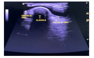
This is the case of a 28 year old male with biopsy proven IgA nephropathy who had received multiple sessions of hemodialysis for the past few months. For the purpose of hemodialysis, a left sided brachio-cephalic AVF was created in the antecubital fossa. Patient presented to us for Doppler evaluation of AVF to assess its maturation. While assessing the left cephalic vein, its course was normal up to the deltopectoral groove. Instead of draining into axillary vein it was seen ascending upwards, superficially crossing the clavicle (Figure 1) and draining into left external jugular vein (Figure 2). Similar course of cephalic vein was seen on right side (Figure 2). The AVF site was patent and was showing a flow rate of 1213 ml/min suggestive of a maturated AVF. CT scan of the patient was not performed in view of deteriorating renal functions and a previous episode of contrast reaction to iodinated contrast.
Discussion
Cephalic vein is a superficial vein of upper limb which develops from the pre-axial vein during embryonic development[2]. The cephalic vein courses in the upper arm lateral to biceps, to the delto-pectoral groove and perforates the clavi-pectoral fascia to drain into the axillary vein [1]. Cephalic vein is used for many clinical and diagnostic procedures. It is used for AVF formation for the purpose of hemodialysis in patients with chronic kidney disease. The cut-down of cephalic vein in the deltopectoral groove is preferred when superior venacaval infusion is necessary. It can also be used for cardiac catheterization. It is a reliable site for cannulation when it is difficult to get access to other veins [3]. Cephalic veins exhibit a wide range of developmental variations in terms of formation, course, and termination [5]. Supraclavicular course of cephalic vein is very rare and available reports indicate that it occurs only in 0.2% cases [6]. In such rare cases, it crosses superficial to the clavicle and terminates into external jugular vein. Supraclavicular course of cephalic vein increases the risk of complications during the clavicular fractures, cephalic vein catheterization, or head and neck surgery. Knowledge of this supraclavicular course of cephalic vein plays an important role during cardiac catheterization, treating clavicle fractures and performing head and neck surgery, to avoid injury to this vascular structure [7].
Conclusion
Supraclavicular course of cephalic vein is a rare variation. This variant course of cephalic vein increases the risk of complications during the clavicular fractures, cephalic vein catheterization, or head and neck surgery. It is a preferred vein for suitable central venous access, AVF formation and pacemaker and defibrillator implantation. Knowledge of such variations of cephalic vein is important for surgeons, clinicians and radiologists as the vein is frequently used for many clinical and diagnostic procedures.
References
- Loukas M, Myers CS, Wartmann CT, Tubbs RS, Judge T, et al. The clinical anatomy of the cephalic vein in the deltopectoral triangle. Folia Morphol (Warsz). 2008; 67: 72–77.
- Standring S. Gray’s anatomy: The anatomical basis of clinical practice. 40th ed. Philadelphia, PA: Churchill Livingstone Elsevier. 2011; pp. 777–822. pp. 899–906.
- Matsuo M, Honma S, Sonomura T, Yamazaki M. Clinical anatomy of the cephalic vein for safe performance of venipuncture. JA Clin Rep. 2017; 3.
- Świȩtoń EB, Steckiewicz R, Grabowski M, Stolarz P. Selected clinical challenges of a supraclavicular cephalic vein in cardiac implantable electronic device implantation. Folia Morphologica (Poland). 2016; 75: 376–381.
- T. Ghosa, Begum S, Roy T, Guptata I. A Rare Variation of Superficial Venous Drainage Pattern of Neck. International Journal of Anatomy, Radiology and Surgery. 2014; 3: 1–3.
- De Maria E, Cappelli S. Cephalic vein with a supraclavicular course: Rare, but do not forget it exists! J Cardiovasc Med (Hagerstown).
- Wysiadecki G, Polguj M, Topol M. Persistent jugulocephalic vein: Case report including commentaries on distribution of valves, blood flow direction and embryology. Folia Morphol (Warsz).