
Journal of Clinical Images and Medical Case Reports
ISSN 2766-7820
Short Report - Open Access, Volume 3
Metastatic spindle cell sarcoma: A rare type of undifferentiated soft tissue neoplasm
Ana Luísa Nunes1*; Ana Lai2; Rui Caetano Oliveira2; Jandira Lima1; Rui Marques Santos1
1 Internal Medicine Department, Centro Hospitalar e Universitário de Coimbra EPE, Portugal.
2 Anatomical Pathology Department, Centro Hospitalar e Universitário de Coimbra EPE, Portugal.
*Corresponding Author: Ana Luísa Nunes
Internal Medicine Department, Centro Hospitalar e
Universitário de Coimbra EPE, Portugal.
Email: ana.luisa.s.nunes@gmail.com
Received : Dec 22, 2021
Accepted : Jan 18, 2022
Published : Jan 25, 2022
Archived : www.jcimcr.org
Copyright : © Nunes AL (2022).
Abstract
Sarcomas are rare and characterised by an incidence of approximately 5 cases/100.000 people. Primary spindle cell sarcomas are a subclassification of soft tissue sarcomas, known as uncertain differentiation tumours, based on their unspecific line of differentiation. It might begin in layers of connective tissue under the skin, between muscles or surrounding organs. Diagnosis can be challenging and delayed due to the often-painless nature of these cancers and the common assumption of an underlying benign condition. The presence of distant metastatic disease at the time of the initial diagnosis is not frequent, but is more likely in large, deep and high-grade malignancy sarcomas. Our case describes an elderly patient diagnosed with an asymptomatic undifferentiated spindle cell sarcoma in the lower limb that developed metastatic disease after intentional curative therapy.
Keywords: CT/MRI LI-RADS; CEUS LI-RADS; gadolinium contrast; MRI contrast agents; congenital heart disease; contrast enhanced ultrasound.
Citation: spindle cell sarcoma; uncertain differentiation sarcoma; soft tissue neoplasms; lung metastasis.
Description
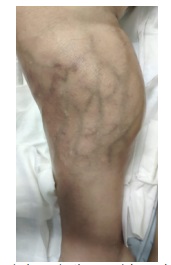
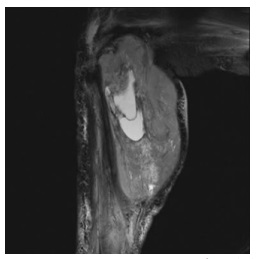
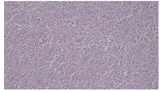
A 79-year-old female patient presented with an asymptomatic and newly-diagnosed severe acute normocytic anaemia. Despite a chronic well-controlled high blood pressure, no other medical history was relevant. On physical examination, a painless enlargement of the posterior compartment of the right lower limb was palpable, with evident collateral circulation (Figure 1). A magnetic resonance imaging was performed, showing a 13 X 12 X 23 cm heterogeneous mass, deeply localized into the gastrocnemius, with significant haemorrhagic component (Figure 2). The 2-[18F]-fluoro-2-deoxy-D-glucose positron emission tomography did not demonstrate any other hypermetabolic lesions. A biopsy approach was performed but the histologic result was compatible with a high-grade sarcoma, without being possible to make a more accurate diagnosis. The patient underwent an above-the-knee amputation of the right lower limb, as curative treatment. The post-operative histological result revealed a spindle sarcoma with aberrant mitotic figures. The immunohistochemical markers were negative for muscle, neural or melanocytic differentiation and the index proliferation marker Ki-67 was more than 40%. The resection margins were tumour free. These findings were suggestive of a completely resected spindle cell sarcoma (Figure 3). After eight months of follow-up, the patient was readmitted due to progressive dyspnoea, with the need for oxygen supply. A chest computed tomography scan showed multiple lung masses, latter confirmed to be lung metastasis. Despite the therapeutic efforts, the patient died of respiratory failure.
Primary spindle cell sarcomas are rare and constitute a subtype of uncertain differentiation soft tissue sarcomas, that lack a specific line of differentiation [1,2]. Delay in diagnosis is common, due to the painless nature of the neoplasm, favouring metastatic disease, that mainly occurs in the lung. Approximately 25% of patients develop distant metastasis after successful treatment of their primary tumour. This incidence increases when tumours present with more than 5 cm in size and deep to the fascia [3].
Declarations
Acknowledgment: None to declare.
Financial disclosure or funding: None to declare.
Conflict of Interest: None to declare.
Author contributions: Ana Luísa Nunes and Ana Lai contributed to the writing of this manuscript; all the authors were involved in the patient’s diagnosis and in the review of this report.
Data availability: The data supporting the findings of this study are available from the corresponding author upon reasonable request.
References
- Sbaraglia M, Bellan E, Dei Tos AP. The 2020 WHO Classification of Soft Tissue Tumours: news and perspectives. Pathologica. 2021; 113: 70-84.
- Feng L, Wang M, Yibulayin F, Zhang H, Yang YL, Ren F, et al. Spindle cell sarcoma: a SEER population-based analysis. Sci Rep. 2018; 8: 5024.
- Allen AH, Gullixson AC. Spindle Cell Sarcoma of the Paraspinal Musculature with Late Pulmonary Metastases. Am J Case Rep. 2019; 20: 828-832.