
Journal of Clinical Images and Medical Case Reports
ISSN 2766-7820
Clinical Image - Open Access, Volume 3
Severe polyostotic fibrous dysplasia presenting as a leg length discrepancy
Vij N1; Belthur MV2*
1University of Arizona College of Medicine, 475 N 5th St., Phoenix, AZ 85004, USA.
2Phoenix Children’s Hospital, Department of Orthopedics, Main Building, Clinic B, 1919 E Thomas Rd, Phoenix, AZ 85016, USA.
*Corresponding Author: Mohan V Belthur
Phoenix Children’s Hospital, Department of
Orthopedics, Main Building, Clinic B, 1919 E Thomas
Rd, Phoenix, AZ 85016, USA.
Tel: 602-934-1660, Fax: 602-933-5245;
Email: mvbelthur@yahoo.com
Received : Dec 14, 2021
Accepted : Jan 21, 2022
Published : Jan 28, 2022
Archived : www.jcimcr.org
Copyright : © Belthur MV (2022)
Keywords: skeletal dysplasias; bone tumors; bisphosphonate therapy; bone grafting.
Citation: Vij N, Belthur MV. Severe polyostotic fibrous dysplasia presenting as a leg length discrepancy. J Clin Images Med Case Rep. 2022; 3(1): 1622.
Case description
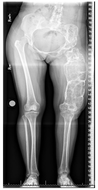
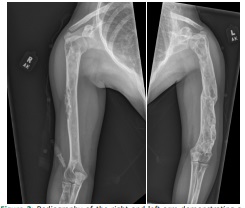
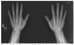
A 16 year old female, recent refugee, presented to the clinic with the complaint of difficulty walking. The patient had been told she had osteoporosis as a child, though did not recollect a formal diagnosis. On physical exam, the patient had good range of motion of the hips, knees, ankles, and feet; however, restricted hip abduction on the left and limited hip internal rotation were noted. The patient walked with a heel-toe gait, a short leg gait on the left, a lateral trunk lean, and a left external foot progression angle. A leg length discrepancy was measured clinically at 4 centimeters, left shorter than right. Radiography of the lower extremities revealed a large expansile lytic lesion throughout the length of the left femur (Figure 1). Radiography of the upper extremities and hands revealed similar lesions (Figure 2 and Figure 3). Genetic analysis was pursued which revealed 5 variants in 5 genes of unknown significance.
The patient was diagnosed with polyostotic fibrous dysplasia based on clinical and radiographic features of the disease. At this point, the decision was made to manage the patient conservatively. A shoe lift was given which allowed the patient to walk comfortably. Given the absence of pain and the relief of her priorly unstable gait, the patient was not interested in a surgical treatment. This surprising case underscores a few interesting point regarding polyostotic fibrous dysplasia. Firstly, this case highlights the painless presentation of radiographically noteworthy polyostotic fibrous dysplasia, which was found incidentally during the work up of a leg length discrepancy [1]. Secondly, bisphosphonate therapy is indicated only in symptomatic polyostotic fibrous dysplasia as it may decrease pain and fracture risk [2], though there is not evidence to support its use in all patients. Lastly, this case highlights the importance of medical screening in refugees [3].
Gene |
Variant |
Zygosity |
Variant classification |
HSPG2 |
c.11920G>A (p.Gly3974Arg) |
heterozygous |
Uncertain significance |
MAP3K7 |
c.1217C>G (p.Ser406Cys) |
heterozygous |
Uncertain significance |
MYO18B |
c.4768G>A (p.Val1590Ile) |
heterozygous |
Uncertain significance |
NOTCH2 |
c.6985C>T (p.Pro2329Ser) |
heterozygous |
Uncertain significance |
POLR1A |
c.4661A>G (p.Tyr1554Cys) |
heterozygous |
Uncertain significance |
Final diagnosis: Polyostotic fibrous dysplasia
Three differential diagnosis:
1. Enchondroma (Ollier’s Disease)
2. Osteochondroma (Multiple Hereditary Exostosis)
3. Non-ossifying fibroma (Jaffe-Campanacci syndrome)
References
- DiCaprio MR, Enneking WF. Fibrous dysplasia: Pathophysiology, evaluation, and treatment. Journal of Bone and Joint Surgery - Series A. 2005; 87.
- Rastogi A, Bhadada SK, Bhansali A. Recurrent femur neck fracture and response to bisphosphonates in polyostotic fibrous dysplasia. Indian J Pediatr. 2012; 79(5).
- The Centers for Disease Control and Prevention. Guidelines for the U.S. Domestic Medical Examination for Newly Arriving Refugees. Immigrant and Refugee Health. 2020.