
Journal of Clinical Images and Medical Case Reports
ISSN 2766-7820
Clinical Image - Open Access, Volume 3
Extended spinal dysraphism: Rachischisis or myeloschisis
Oze Koudouhonon Rita; Yehouenou Tessi Romeo Thierry*; El Haddad Siham; Allali Nazik; Chat Latifa
Radiology Department, Ibn Sina Paediatric Teaching Hospital, Mohammed V University, Rabat, Morocco
*Corresponding Author: Yehouenou Tessi Romeo Thierry
Department of Radiology, Ibn Sina Paediatric Teaching
Hospital, Mohammed V University, Rabat, Morocco.
Tel: +212-613-211-125;
Email: nactessi@yahoo.fr
Received : Dec 06, 2021
Accepted : Jan 24, 2022
Published : Jan 31, 2022
Archived : www.jcimcr.org
Copyright : © Thierry YTR (2022).
Citation: Oze KR, Yehouenou Tessi RT, El Haddad S, Allali N, Chat L. Extended spinal dysraphism: Rachischisis or myeloschisis. J Clin Images Med Case Rep. 2022; 3(1): 1627
Description
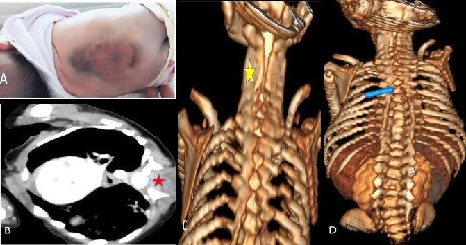
Myeloschisis (rachischisis) is a rare congenital malformation linked to incomplete development of the spine and characterized by posterior opening of the vertebrae [1,2]. It is spread over more than one floor or even the entire spine and incompatible with life. Its diagnosis is clinical, confirmed by ultrasound and cross-sectional imaging [2]. This anomaly in its thoracic location is rare. There are two types: Segmentation and development defects [4]. Developmental defects include hemi vertebrae and failure of neural tube fusion which in turn includes the simple cleft of the posterior arch of the vertebrae (spina bifida) [3]. It can involve one or more vertebral arches, determining extensive spinal dysraphism. Certain factors favor their appearance. According to the literature, only one article published in 1972 by Levy et al, which reported an incidence of 0.01% of thoracic spina bifida [3]. We report the case of a 33-day-old female infant born at term in breech presentation, with a pregnancy that was not well followed. There was a notion of taking fenugreek during pregnancy. Hospitalized for meningitis, with a dysmorphic face, clinically presented a dorsolumbar spina bifida (Figure 1). She had received antibiotic therapy and regular dressings since her admission. A cerebral-medullary scan was performed and showed a progressive opening of the spinous processes from the cervical and dorsal spine, more marked at the height of the spinous process of the fourth dorsal vertebra to distality, with issue of meningeal structures and spinal cord.
Conflicts of interest: The authors declare that they have no conflicts of interest.
References
- Manenti G, Iundusi R, Picchi E, Marsico S, D’Onofrio A, Rossi G, et al. Anatomical variation: T1 spina bifida occulta. Radiological findings. Radiol Case Rep. 2016; 12: 207-209.
- Jaganmohan D, Subramaniam P, Krishnan N, Mahajan P. Two cases of craniospinal rachischisis totalis: Role of magnetic resonance imaging in diagnosis and review of neural tube defects in the Indian context with implications for folate fortification. Journal of pediatric neurosciences. 2017; 12: 32-35.
- Manenti G, Iundusi R, Picchi E, Marsico S, D’Onofrio A, et al. Anatomical variation: T1 spina bifida occulta. Radiological findings. Radiology case reports. 2017; 12: 207-209.