
Journal of Clinical Images and Medical Case Reports
ISSN 2766-7820
Case Report - Open Access, Volume 3
Conservative management for fetal megacystis: A clinical based approach
Lisa Hassan1; Richard Kiritta1*; Clotrida Chuma1; Edgar Ndaboine1; Adolphine Hokororo2; Albert Kihunrwa1
1 Department of Obstetrics and Gynecology, Bugando Medical Centre, Catholic University of Health and Allied Sciences, Tanzania.
2 Department of Paediatrics and Child Health, Bugando Medical Centre, Catholic University of Health and Allied Sciences, Tanzania.
*Corresponding Author: Richard Kiritta
Department of Obstetrics and Gynecology, Bugando
Medical Centre, Catholic University of Health and
Allied Sciences, Mwanza, Tanzania.
Email: kiritta2002@yahoo.com
Received : Jan 13, 2022
Accepted : Feb 02, 2022
Published : Feb 09, 2022
Archived : www.jcimcr.org
Copyright : © Kiritta R (2022).
Abstract
Background: Fetal megacystis is an enlargement of the fetal urinary bladder that is a rare ultrasonography finding during routine antenatal screening. Though intrauterine genetic screening is widely used to determine the prognosis of this condition in resource rich settings, in the developing world, clinician often has to rely on clinical judgement for proper counselling and management of women with such foetal presentations.
Case presentation: We present a case of a 31 years old lady who presented at our facility as a referral case at a gestational age of 25 weeks for termination of pregnancy due to an incidental ultrasonography finding of an enlarged fetal urinary bladder with oligohydramnious. Initial evaluation at our facility led to diagnosis of foetal megacystis with no other congenital anomalies. An informed expectant management was carried out under close follow up and finally delivered a 4.2 kg baby by caesarean section at 39 weeks. The baby is currently 4months of age, with normal renal function parameters on routine care and urological follow up.
Conclusion: Foetal megacystis is a rare condition with diverse aetiology. Genetic screening for chromosomal anomalies is a major prognostic indicator however in resource limited setup, a clinician has to rely on clinical judgement to provide the patient with the necessary information to make an informed decision between expectant management or termination of pregnancy.
Keywords: megacystis; oligohydramnios; urinary bladder; genetic screening; case report.
Citation: Hassan L, Kiritta R, Chuma C, Ndaboine E, Hokororo A, et al. Conservative management for fetal megacystis: A clinical based approach. J Clin Images Med Case Rep. 2022; 3(2): 1647.
Introduction
Fetal megacystis is an enlargement of foetal bladder in utero, a diagnosis that is usually made when a pre-natal ultrasonography urinary bladder longitudinal diameter is more than 7 milimetres. The Aetiology of foetal megacystis is diverse and is directly related to the prognosis of the condition. In resource limited setup, knowing the exact cause for the condition is often difficult due to limitations in diagnostic modalities and genetic screening in particular, hence the decision for expectant management or pregnancy termination relies on clinicians’ judgement and close follow up on clinical parameters. We present a case of distended fetal bladder at gestation of 25 weeks that was managed expectantly to birth, describing possible causes, clinical presentation, diagnosis modalities, predictors of outcome and management of such patients.
Case presentation
We present a 31 years old lady who came to our facility while pregnant at a gestation age of 25 weeks after being referred from a lower health facility due to an abnormal obstetric ultrasound finding at the primary site warranting termination of pregnancy. This was a coincidental finding on a routine ultrasound scan during her routine antenatal clinic (ANC) visit. She was otherwise well and no any other pregnancy complications.
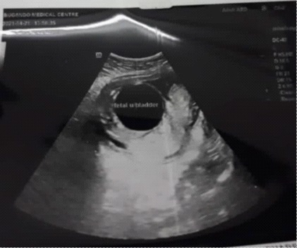
On the day of an outpatient visit, she was physically well. Her antenatal card showed she had attended three [3] visits, she was HIV negative, and tested negative for sexually transmitted infections and had all the required ANC essentials as per schedule. Physical examination revealed a middle-aged lady, blood pressure of 124/80 mmhg. Abdominal examination showed a gravid abdomen of 20 cm fundal height, non-tender and no organomegaly. Blood work showed normal haemoglobin level of 12 g/dl and VDRL was non-reactive. A repeat obstetric scan was done and revealed a live intrauterine pregnancy at gestation age of 18 weeks with a fundal posterior placenta, a distended foetal urinary bladder and oligohydramnios (Figure 1), no other foetal anomalies were detected. An informed decision for expectant management was made by the patient, and was kept on close foetal surveillance at our facility. Integrated iron supplements, malaria prophylaxis and deworming were done as per routine ANC care and was scheduled for a repeat ultrasound scan after 5 weeks. At 23 weeks of gestation an obstetric scan was done and showed a live intrauterine pregnancy at a gestation age of 23 weeks with estimated foetal weight of 643 grams, the amniotic fluid was found to be adequate with deepest vertical pocket measuring 9 centimetres and no other fetal anomalies noted. Follow up visits were done monthly apart together with obstetric scans which were all normal. At gestational age of 39 weeks and 4 days, an elective caesarean section was done due to suspected big baby of whom she delivered a 4.2 kg male baby with no complications and no physical congenital malformations.
An initial paediatrician review and sonographic evaluation of the baby revealed a normal echocardiography; ordered abdominal pelvic ultrasound was less informative and hence a CT scan of the abdomen and pelvis was done which revealed a thickened bladder wall and hyperdense right kidney. The renal function tests were slight elevated with serum creatinine of 183 umol/l(normal values 62-106 umol/l), however a repeat Renal function test 2 weeks later showed a significant decline to a normal value of 85 umol/l.
Subsequent follow up of the baby at paediatric and urology clinic revealed a baby was fairing well with a normal growth curve, normal urine output and normal sonographic findings at 3 months of age.
Discussion
Foetal megacystis is the distension of the foetal urinary bladder diagnosed by ultrasonographic longitudinal bladder diameter of more than 7 millimetres. It occurs in about 0.06% of pregnancies in the first trimester with the actual prevalence remaining unknown beyond this trimester [1]. Deranged renal function after birth and perinatal mortality encompass this diagnosis [2]. In our case like other reported cases, foetal megacystic was incidentally detected via a routine second trimester obstetric ultrasound [3].
Megacystis have been associated with posterior urethral valves(57%), urethral atresia/stenosis (7%) and karyotype anomalies (15%) including trisomy 18 and 21 [4]. These may evolve to spontaneous resolution or progress to obstructive uropathy [3]. Poor prognosis has been associated with chromosomal anomalies and genetic screening is widely used as a prognostic indicator and in counselling and management of such cases in the developed world. On clinical grounds, urethral atresia/stenosis has been associated with poor outcome, however in our patients’ case, the resolution of oligohydramnious seen on serial obstetric scans helped the clinician to rule out the possibility of urethral atresia/stenosis hence offered the possibility of a better prognosis.
Few cases of isolated congenital foetal mega cystic without complications have been reported [5,6]. Our infant initially presented with renal function derangements, thickened bladder wall and left renal agenesis few weeks after birth with no other visible congenital anomalies noted both on abdominal CT scan and echocardiogram. Failure to establish karyotype abnormalities in our patients’ case due to resource limitations made us to continue with expectant management relying on clinical judgement with follow up on renal function tests and repeat ultrasound scans which showed improvement over time.
Counselling on termination of pregnancy or expectant management still remains a challenge in managing this rare case. Few literatures have described some prognostic factors for fetal outcome which may influence the course of pregnancy. Among the predictors of outcome include a proper sonography to exclude any renal damage (dysplasia/renal cysts), serial amniotic fluid measurements, presence of other malformations and fetal sex [7,8]. Absence of congenital anomalies on the initial obstetric ultrasound and subsequent spontaneous correction of amniotic fluid volume were the main prognostic factors that made clinicians to expect good outcome in our patients’ case.
A meta-analysis review revealed that some of the foetus with megacystis can have a good prognosis and therefore a good outcome. From this study oligohydromnious and gestation age at diagnosis were superior in predicting survival. Normal AFI showed good functioning renal system and a good pulmonary function [9-11]. This was our basis for expectant management as our AFI significantly improved four weeks later up to birth. Increased gestational age at diagnosis still remains as high as AFI among the major predictors of survival rates, being that diagnosis at second and third trimester had good outcomes and our patient was diagnosed at the second trimester.
Several authors recommend future control trials for assessment of the feasibility of intrauterine intervention of foetus with megacystis. Some documented intervention includes vesicoscentesis and vesicoamniotic shunt done intrauterine. However no significant future survival rate and renal function difference has been observed between foetus who underwent these interventions to those who were managed conservatively [12,13]. In our case at 4 months of age with normal renal function test parameters and still under conservative care and close follow up by the team with serial renal function evaluation, we believe careful selection of patient with close follow up remains the standard of care in resource limited setup. To our knowledge, the longest follow up case of foetal megacystis is 10 years who had no complications and neither surgical interventions among the very rare cases who have been followed up without any complication [5]. Therefore we believe this will be another case that we will follow up in the long run.
Conclusion
Foetal megacystis is a rare incidental finding in routine Obstetric ultrasound. Decision for expectant management or termination of pregnancy in resource limited setup should rely on clinical judgement and close follow up of selected cases. An informed decision for conservative management of cases is mandatory due to the diversity in etiological causes that perhaps cannot be determined in resource limited setup.
Declarations
Patient’s perspective: The care provide to the patient was free of charge and timely with a follow up plan.
Acknowledgement: We are grateful to the Departments of Obstetrics and gynecology, paediatrics, radiology and urology at Bugando Medical Centre.
Timeline: Patient was first seen at our clinic on May 2021, delivered on September 2021 by caesarean section, the baby is currently on his 4th month follow up with the urology clinic.
Author’s contribution: LH and RK prepared this case report. The other co-authors contributed to the management and writing of the case report.
Funding: The cost of care to this patient was waived by the hospital as per Government protocol. The cost of preparing the manuscript and publication was covered by the authors andDirectorate of research and publications-Catholic University of Health and Allied Sciences.
Ethical approval, consent to participate and publish: Written informed consent was provided by the patient for publication of this case report and related images. Additional consent was thought and granted by the joint CUHAS/BMC research and ethical committee. All the relevant copies are available for review by the Editor in Chief of this journal.
References
- Sebire NJ, Von Kaisenberg C, Rubio C, Snijders RJM, Nicolaides KH. Fetal megacystis at 10-14 weeks of gestation. Ultrasound in Obstetrics and Gynecology. 1996; 8: 387-90.
- Fontanella F, Duin L, Pajkrt E, Bekker M, Bax CJ, Bilardo CM, et al. Antenatal Workup of Early Megacystis and Selection of Candidates for Fetal Therapy. 2018; 1-7.
- Fontanella F, Adama PN, Scheltema V. Antenatal Workup of Early Megacystis and Selection of Candidates for Fetal Therapy. 2018; 1-7.
- Taghavi K, Sharpe C, Stringer MD. SC. J Pediatr Urol [Internet]. 2016; Available from: http://dx.doi.org/10.1016/j.jpurol.2016.09.003
- Nakamura T, Neonatology D, Metropolitan T, Children H. Isolated congenital megacystis with spontaneous resolution : a case report. 2017; 63(2): 116-20.
- Johnson EK, Nelson CP. Spontaneous resolution of isolated congenital megacystis: The incredible shrinking bladder. J Pediatr Urol [Internet]. 2013; 9(1): e46-50. Available from: http:// dx.doi.org/10.1016/j.jpurol.2012.07.012
- Pellegrino M, Visconti D, Catania VD, D’Oria L, Manzoni C, Grella MG, et al. Prenatal detection of megacystis: not always an adverse prognostic factor. Experience in 25 consecutive cases in a tertiary referral center, with complete neonatal outcome and follow-up. J Pediatr Urol [Internet]. 2017; 13(5): 486.e1-486.e10. Available from: http://dx.doi.org/10.1016/j.jpurol.2017.04.001
- Fievet L, Faure A, Coze S, Harper L, Panait N, Braunstein D, et al. Fetal megacystis: Etiologies, management, and outcome according to the trimester. Urology. 2014; 84(1): 185-90.
- Chen L, Guan J, Gu H, Zhang M. Outcomes in fetuses diagnosed with megacystis: Systematic review and meta-analysis. Eur J Obstet Gynecol Reprod Biol [Internet]. 2019; 233: 120-6. Available from: https://doi.org/10.1016/j.ejogrb.2018.12.007
- Robyr R, Benachi A, Daikha-Dahmane F, Martinovich J, Dumez Y, Ville Y. Correlation between ultrasound and anatomical findings in fetuses with lower urinary tract obstruction in the first half of pregnancy. Ultrasound Obstet Gynecol. 2005; 25(5): 478-82.
- Van Mieghem T, Ryan G. The PLUTO trial: A missed opportunity. Lancet [Internet]. 2013; 382(9903): 1471-3. Available from: http://dx.doi.org/10.1016/S0140-6736(13)61446-4
- Morris RK, Malin GL, Khan KS, Kilby MD. Systematic review of the effectiveness of antenatal intervention for the treatment of congenital lower urinary tract obstruction. BJOG An Int J Obstet Gynaecol. 2010; 117(4): 382-90.
- Ruano R, Sananes N, Sangi-Haghpeykar H, Hernandez-Ruano S, Moog R, Becmeur F, et al. Fetal intervention for severe lower urinary tract obstruction: A multicenter case-control study comparing fetal cystoscopy with vesicoamniotic shunting. Ultrasound Obstet Gynecol. 2015; 45(4): 452-8.