
Journal of Clinical Images and Medical Case Reports
ISSN 2766-7820
Case Report - Open Access, Volume 3
Acute necrotizing encephalopathy associated with COVID-19 infection in a child
Erdal Karavas1; Oğuzhan Tokur2*; İsmail Topal3 ; Sonay Aydın1
1 Erzincan Binali Yildırım University, Department of Radiology, Erzincan, Turkey.
2 Ankara Training and Research Hospital, Department of Radiology, Ankara, Turkey
3 Erzincan Binali Yildırım University, Department of Pediatrics, Erzincan, Turkey.
*Corresponding Author: Oguzhan Tokur
Department of Radiology, Ankara Training and
Research Hospital
Email: oguzhantokur@gmail.com
Received : Dec 20, 2021
Accepted : Feb 07, 2022
Published : Feb 14, 2022
Archived : www.jcimcr.org
Copyright : © Tokur O (2022).
Abstract
Every day, new knowledge on the SARS-CoV-2 epidemic spreads throughout the globe. It presents typically with fever and respiratuar symptoms like shortness of breath and cough but neurologic complications aren’t still well known. Although neurologic complications of SARS CoV-2 infection have been reported, particularly in adults, rarely reported in children. Acute Necrotic Encephalopathy (ANE) is one of the rare neurological complications associated with coronavirus disease 2019 (COVID-19) and usually has poor prognosis in pediatric population. In this report, we present a rare case of acute necrotic encephalopathy due to COVID-19 infection in a 2-year-old child. To our knowledge, there has been limited reports of acute necrotic encephalopathy associated with COVID-19 in children in the literature.
Keywords: Covid-19; SARS CoV-2; pediatric; necroziting encephalopathy.
Citation: Karavas E, Tokur O, Topal I, Aydın S. Acute necrotizing encephalopathy associated with COVID-19 infection in a child. J Clin Images Med Case Rep. 2022; 3(2): 1657.
Introduction
The COVID-19 pandemic caused by the SARS-CoV-2 virus has the potential to be fatal, particularly in adults all over the world. Although the typical clinical presentation of the infection is fever, weakness and respiratory system symptoms such as cough and shortness of breath, neurological manifestations may occur. Adults may develop ischemia and intracerebral hemorrhage as a result of SARS-CoV-2 infection, according to the literature [2]. In contrast, neurological impairment in children has been reported infrequently [2,6,7]. We present a rare case of Acute Necrotic Encephalopathy (ANE) caused by SARS-CoV-2 infection in a 2-year-old child.
Case presentation
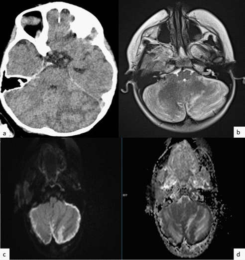
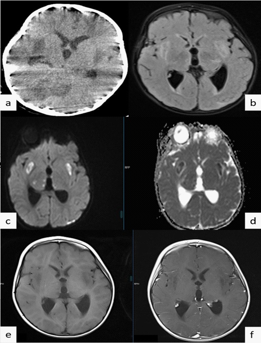
A 2-year-old girl presented the emergency room with weakness and a high fever after five days her father was diagnosed with COVID-19. Her medical history was unremarkable. Except for her body temperature of 38.5 degrees Celsius, her physical examination was unremarkable. Because his father had a history of COVID-19, she was tested with a nasal swab PCR and diagnosed with COVID-19. She was admitted to the pediatric unit for observation and treatment. On the fourth day of follow-up, non-contrast brain Computed Tomography (CT) and Magnetic Resonance Imaging (MRI) were performed after a tendency to sleep was observed. She also had seizures of licking, smacking, and swallowing. On the CT images, there were scattered hypodense areas in the cerebellum and bilateral thalamus (Figure 1a and 2a). T2-weighted (T2WI) (Figure 1b) and fluid-attenuated inversion recovery (FLAIR) images (Figure 2b) from MRI revealed hyperintense areas in the same locations as CT. The cerebellum and bilateral thalamus showed restricted diffusion on Diffusion Weighted Images
(DWI) (Figure 1 and 2). Bilateral thalamic peripheral rim enhancement was also observed. (Figures 2e and f). Polymerase Chain Reaction (PCR)–based testing of Cerebrospinal Fluid (CSF) revealed herpes simplex virus 1 and 2, varicella zoster and SARS CoV-2 virus were negative. But she was diagnosed with acute necrotic encephalopathy with clinic and imaging features.
Discussion
Although COVID-19 infection is typically identified by respiratory symptoms, it can also result in neurological complications [1]. The rise in the number of cases during the pandemic has revealed the need to learn more about neurological complications in children and adults. However, there is still a scarcity of information about neurological complications, particularly in children [2]. There are some cases of COVID-19-related ANE in the literature, but the majority of them are in adults [1,3-5]. To the best of our knowledge, there have been only a few reports of acute necrotic encephalopathy (ANE) in children associated with COVID-19 in the literature [6,7].
Acute Necrotic Encephalopathy (ANE) is a relatively uncommon neurological complication of SARS Cov-2 infection [6]. The etiology and pathophysiology are not well understood [8]. It has been proposed in several hypotheses that it may develop as a result of cytokine storm or virus-induced endothelial damage to the blood-brain barrier [1,6].
Seizures, vomiting, and loss of consciousness are common symptoms of ANE after 12-72 hours of viral infection. These symptoms, however, are not unique to ANE. Some cases in the literature revealed that PCR assays can only rarely detect pathogens in CSF [9]. As a result, neuroimaging plays an important role in diagnosis. Bilateral distributed hypoattenuation on brain CT and T2WI/FLAIR hyperintensities on MRI in the cortex, periventricular white matter, thalami, basal ganglia, brain stem, and cerebellar hemispheres are typical findings. Restricted diffusion can be seen on Diffusion Weighted Imaging (DWI) in the same areas, which is a sign of necrosis. Contrast-enhanced images may show peripheral rim enhancement. Susceptibility-Weighted Images (SWI) can also show whether there is a hemorrhagic component [1,4,5].
The first option in the differential diagnosis is Acute Disseminated Encephalomyelitis (ADEM). It results in asymmetric and ill-defined lesions in the periventricular and basal ganglia, whereas ANE results in symmetric, well-defined lesions, particularly in the bilateral thalamus [4].
There is no specific treatment for COVID-associated acute necrotic encephalopathy at the moment. High-dose corticosteroids, polyvalent immunoglobulins or plasmapheresis can be used to treat the condition, but they will be more effective if diagnosed and treated early [3,4,6].
Despite the fact that ANE is rarely reported, as in this case, it is important to remember it as a possible complication in the pediatric population to allow for prompt diagnosis and treatment.
All procedures performed in studies involving human participants were in accordance with the ethical standards of the institutional and/or national research committee and with the 1964 Helsinki declaration and its later amendments or comparable ethical standards. Written consent was acquired from the child’s parents.
Declarations
Patient consent: The authors certify that they have obtained all appropriate patient consent forms. In the form, the legal guardian has given his consent for images and other clinical information to be reported in the journal. The guardian under stands that names and initials will not be published and due efforts will be made to conceal patient identity, but anonymity cannot be guaranteed.
Competing interest: The authors declare that they have no competing interests.
Availability of data and material: The data sets used and/ or analysed during the current study available from the corresponding author on reasonable request.
Authors’ contributions: This study was directed and coordinated by OT and SA as the principal investigator, provided conceptual and technical guidance for all aspects of the project. OT, SA, EF and FDG planned and performed the analysis of the MRI and CT images. Literature search was suggested and executed by OT and FDG. Design of the study was by OT and SA. The manuscript was written by OT and commented on by all authors. The authors read and approved the final manuscript
Ethical approval and consent to participate: Not applicable.
Consent for publication: Yes, written consent to publish this information was obtained from study participant.
Acknowledgements: Not Applicable.
References
- Mullaguri N, Sivakumar S, Battineni A, Anand S, Vanderwerf J. COVID-19 Related Acute Hemorrhagic Necrotizing Encephalitis: A Report of Two Cases and Literature Review. Cureus. 2021; 13: e14236.
- Lindan CE, Mankad K, Ram D, Kociolek LK, Silvera VM, et al. Neuroimaging manifestations in children with SARS-CoV-2 infection: A multinational, multicentre collaborative study. The Lancet Child & Adolescent Health. 2021; 5: 167-177.
- Kumar N, Kumar S, Kumar A, Pati BK, Kumar A, et al. Acute necrotizing encephalitis as a probable association of covid-19. Indian Journal of Critical Care Medicine: Peer-reviewed, Official Publication of Indian Society of Critical Care Medicine. 2020; 24: 991-994.
- Delamarre L, Gollion C, Grouteau G, Rousset D, Jimena G, et al. COVID-19–associated acute necrotising encephalopathy successfully treated with steroids and polyvalent immunoglobulin with unusual IgG targeting the cerebral fibre network. Journal of Neurology, Neurosurgery & Psychiatry. 2020; 91: 1004-1006.
- Poyiadji N, Shahin G, Noujaim D, Stone M, Patel S, et al. COVID-19–associated acute hemorrhagic necrotizing encephalopathy: Imaging features. Radiology. 2020; 296: E119-E120.
- McAbee GN, Brosgol Y, Pavlakis S, Agha R, Gaffoor M, et al. Encephalitis associated with COVID-19 infection in an 11-year-old child. Pediatric neurology. 2020; 109: 94.
- Kinikar A, Kulkarni R, Rajput U, et al. Acute Encephalopathy in a Child with Coronavirus Disease-2019 Infection. Pediatr Inf Dis. 2020; 2: 62–63.
- Gökharman D, Ünlü HA, Aydın S, Tek C, Kosar PN, et al. Acute necrotizing encephalopathy as a H1N1 complication: A rare case. Cumhuriyet Tıp Dergisi. 2017; 39: 696-699.
- Dixon L, Varley J, Gontsarova A, Mallon D, Tona F, et al. COVID19-related acute necrotizing encephalopathy with brain stem involvement in a patient with aplastic anemia. Neurology-Neuroimmunology Neuroinflammation. 2020; 7: e789.