
Journal of Clinical Images and Medical Case Reports
ISSN 2766-7820
Case Report - Open Access, Volume 3
Venous leg ulcers: A practical guide to management
Ayad Jindeel
Essentia Health, Duluth MN, USA.
*Corresponding Author: Ayad Jindeel
Essentia Health, Duluth MN, USA.
Email: aj1med@aol.com
Received : Jan 06, 2022
Accepted : Feb 10, 2022
Published : Feb 17, 2022
Archived : www.jcimcr.org
Copyright : © Jindeel A (2022).
Abstract
Venous leg ulcers are common. They are usually chronic, costly, and recurrent. They adversely impact patients psychologically and physically. Despite the availability of simple, cost-effective measures to heal them and prevent their recurrence, their incidence has not been decreasing. For a variety of reasons like socioeconomic issues, increasing prevalence of obesity and sedentary lifestyle, treatment continues to be challenging. Management of venous leg ulcers start with taking detailed history, doing thorough examination, and sometimes ordering labs and imaging studies. This will enable the providers to make an accurate diagnosis, uncover the underlying pathology and assess any associated conditions like ischemia. The underlying pathology could be abnormal valvular reflux, venous obstruction, or combination of the two. Gravity and intraabdominal pressure are the main forces causing and aggravating venous leg ulcers. It is critical to define the underlying pathology and any associated condition that could interfere with the healing process like obesity, sedentary lifestyle, sleeping in chairs and ischemia. In addition to appropriate wound care, elevation of feet above heart level, wearing knee high compression stockings, doing regular exercises and weight loss in patients who are obese are usually adequate to heal most patients with venous leg ulcers. There is no specific frequency and duration is required for elevating the feet however the more frequent and the longer time the patients elevate their feet, the quicker their ulcers heal. Using effective compression over the wound dressing is important and requires frequent monitoring. Lower extremity exercises are critical even in patients who are not ambulatory. These measures are important even in patients who may need venous procedures to help heal their ulcers and prevent their recurrence.
Keywords: venous ulcer; gravity; obesity; leg elevation; exercise; compression stockings.
Citation: Jindeel A. Venous leg ulcers: A practical guide to management. J Clin Images Med Case Rep. 2022; 3(2): 1670.
Introduction
Venous leg ulcers are common [1-3], costly to treat [4] and usually chronic [5-8]. Only 45% of patients with venous stasis ulcers were found without recurrence after 5 years of follow up [5]. There incidence has not changed despite the availability of simple, cost-effective measures to heal them and prevent their recurrence (Table 2). Treatment continues to be challenging for a variety of reasons like increasing prevalence of obesity and sedentary lifestyle [9] (Table 3).
Case report
A 58-year-old male presented with a left leg ulcer that has been open for 34 years! He has been managed with a variety of wound dressings, compressions, Unna Boot, antibiotics, topical ointments, skin grafting, sclerotherapy, and thermal ablation. He complained of pain, swelling, discoloration of his left leg and profuse drainage from his left leg ulcer. He is now more worried about losing his leg. He worked as a cashier and stands most of his shift. His daily wound dressing and sock are usually soaked after several hours. He changes the dressing before and after work. He sleeps in a bed. He does not elevate his feet. He did not find compression stockings to be helpful, so he stopped using them. He has no other medical problems and takes Ibuprofen for pain. He does not smoke or drink. He lives alone. His mother has varicose veins.
On examination, he appeared anxious. His vitals, heart, lung, and abdominal exams were normal. There were large varicose veins starting in the left upper anteromedial thigh coursing down the medial thigh, knee and calf leading to the area of ulceration. He had swelling, hyperpigmentation, lipodermatosclerosis and a large ulcer on the left medial ankle with dry, scaly, and thickened skin surrounding the ulcer (Figure 1). His pedal pulses are palpable. He was walking with a limp. His left ankle joint range of motion was limited. His venous competence study showed no evidence of left lower extremity DVT. Waveforms from the common femoral vein were spontaneous and phasic with normal augmentation bilaterally. There was severe valvular reflux of the saphenofemoral junction, the great saphenous vein, and related varicosities. He was advised to elevate his feet above heart level as long and as frequently as possible throughout the day, stop using any topical creams or ointments, wash his left leg and the ulcer with warm water and soap prior to applying wound dressing, apply plain Vaseline to the skin around the ulcer and plain foam dressing on the ulcer, wrap with Kerlix then apply ACE bandage or fitted 20-30 mmHg, knee-high compression stockings. He was advised to do regular exercises and to do stretches of his ankle joints to increase their range of motion.
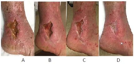
The area of ulceration and the surrounding skin improved quickly (Figure 1). Endovenous Laser ablation of the left great saphenous vein with sclerotherapy of the remaining varicose veins were done to accelerate healing and prevent recurrence. The ulcer healed six weeks after the patient’s first visit. The patient was extremely happy and five years later had no recurrent ulceration.
Discussion
Venous leg ulcer is a clinical diagnosis that start with taking a detailed history, doing a thorough examination, and sometimes ordering labs and imaging studies. This will enable the providers to make an accurate diagnosis, assess any associated conditions like ischemia and uncover the underlying pathology as abnormal valvular reflux, venous obstruction, or a combination of the two.
Patient with a history of lower extremity deep vein thrombosis (DVT) likely has post-phlebitis syndrome. Some medications like calcium channel blockers cause leg swelling. Topical ointments like antibiotics could cause contact dermatitis. Some medications can cause leg ulceration like hydroxyurea. What is their occupation and what are their hobbies? How long do they sit or stand? [8]. Venous insufficiency is worsened in those who sit or stand long hours like truck drivers and barbers. What type of physical activities do they usually do? Do they sleep in a bed or chair? Do they wake up with foot pain when lying down flat suggesting possible ischemia? Do they elevate their feet? Does their leg pain get better or worse after sitting, standing, or walking? Symptoms that improve with lying flat or elevation of feet and worsen with sitting or standing are more likely to be venous, while leg pain triggered by walking is more likely ischemic, neurogenic, or musculoskeletal. Do they live alone? Do they live with someone who is able and available to help with their care? Are there any other family members, friends, neighbors that could help? A major part of the evaluation is to assess if the patient can comply with the treatment plan and to try to problem solve barriers with patients and all those involved in their care. Family history of varicose veins and venous stasis ulceration is common.
Examination: It is valuable to observe patients walking and transferring from chair to exam table. Is the patient able to lie flat on the exam table? If not, why not? Checking vitals, and doing cardiac, pulmonary, and abdominal exams are essential to rule out systemic diseases causing or aggravating venous insufficiency [10,11]. Abdominal examination may reveal abdominal pathology contributing or causing patient’s venous insufficiency like ascites or abdominal tumors. Abdominal wall varicose veins suggest blockage of the abdominal veins (iliac and IVC) or cirrhosis. A positive stemmer test suggests the presence of lymphedema. Femoral bruit suggest the presence of significant arterial disease. The lower extremity exam should include any ulcerations, varicose veins, lipodermatosclerosis, stasis dermatitis, foot deformities, neuro exam, joint exam, and pedal pulses. Sometimes it is challenging to palpate pedal pulses secondary to edema and thickening of the skin or the presence of arterial disease. Portable small Doppler machines are inexpensive, quick, easy to use and very effective in the evaluation of pedal pulses. Absent Doppler signal indicates possible occlusion, while multiphasic signal indicates lack of significant arterial disease. Monophasic doppler signal suggests the presence of arterial disease or significant inflammation. Ankle brachial index (ABI) is an easy and reliable study to assess the presence and severity of arterial disease in most patients. Patients with diabetes or renal insufficiency often have noncompressible vessels, making measurement of ABI not possible or less reliable. A segmental arterial study, arterial duplex, and CT angiogram may be needed to evaluate the lower extremity arterial circulation. Excluding and addressing significant ischemia is crucial in management of leg ulcers.
Venous leg ulcer is a clinical diagnosis and extensive work up is not needed in most cases. It is important to think about the underlying pathology of venous insufficiency as primary varicose veins, post-phlebitis syndrome or compression of veins as in May-thurnner syndrome. The most common differential diagnoses of venous ulcer are listed in (Table 1), [12-14]. Neuropathic ulcers are the most common ulcer distal to the ankle joint while venous ulcers are the most common from the ankle to the knee. Patients could have different types of ulcers on the same leg or the other leg at the same time or different times. They could start with one type of ulcer and then develop another type so regular assessment is crucial during the treatment course.
Table 1:The most common differential diagnoses of venous ulcer.
Common Differential Diagnosis of Venous Ulcer |
1. Neuropathic |
Table 2:Essential elements of the management of Venous leg ulcers.
Essential Elements of the Management of Venous Leg Ulcers |
1. Accurate Diagnosis |
2. Identifying and addressing coexisting diagnoses that impact venous ulcer healing like ischemia. |
3. Measures to reduce leg swelling such as limiting sitting, elevation of feet above heart level, wearing fitted compression stockings and doing regular exercises. |
4. Wearing fitted compression stockings on a regular daily basis to help heal venous ulcers and prevent their recurrence. |
5. Healthy lifestyle changes especially increasing physical activities, weight management and smoking cessation. |
6. Wound care is essential and very effective when done in conjunction with above measures. When wound care fails to heal a venous ulcer, it is usually because patients are not adopting other measures, wrong diagnoses or an unaddressed coexisting medical condition like ischemia. |
7. Venous procedures are indicated in some patients to help heal or prevent recurrent ulcerations. However, they are not a magic wand for every patient. |
Table 3:Challenges in the management of Venous leg ulcers.
Challenges in the Management of Venous Leg Ulcers |
1. The pervasive negative impact of gravity and intraabdominal pressure whenever patients with venous insufficiency have their feet below heart level. There is no pill to counterbalance these forces. Their impact is not limited to a season, location, part of the day, race, gender, or age. Furthermore, more and more people are affected secondary to increasing prevalence of sedentary lifestyle and obesity. |
2. Patients related factors like obesity, sleep apnea, diabetes, neuropathy, back pain, tobacco use, poor compliance, and sedentary lifestyle. In one study, 35% of patients did not have a 10- minute walk once a week. And only 39% Of patients with venous leg ulcers adhered to wearing compression stockings.21 |
3. Socioeconomic factors like loneliness, social isolation, poverty, homelessness, lack of health insurance, inability to afford buying compression stockings. |
4. Healthcare related factors as |
Identification and treatment of coexisting conditions that could interfere with venous ulcer healing is critical like ischemis, obesity and sedentary lifestyle. In patients with significant lower extremity ischemia, healing of the venous ulcer will be slow or will not happen before addressing their ischemia.
Also, in these patients wearing compression stockings and elevation of feet could worsen their ischemia. Revascularization will also help them to comply with elevation of their feet and wearing compression stockings. Patients may have peripheral neuropathy making them more prone to pressure ulcer and repeated trauma. If the patient has obesity, there is nothing more effective to heal their venous ulcers than weight loss [15,16].
Measures to reduce leg swelling and wound discharge: We strongly recommend against sleeping in chair and to limit how long they sit to minimum. We recommend they elevate their feet above heart level as long as tolerated and as frequently as possible throughout the day [17,18]. We encourage them to read, watch TV, and use their phone while lying down with their feet above heart level. We recommend that patients use a foam wedge to elevate their feet. Elevating feet while sitting in a chair is not helpful. Elevation of feet above heart level reduces hydrostatic pressure, filtration of fluids, macromolecules, red blood cells, inflammation and helps drain the accumulated interstitial fluids via lymphatics and veins. It is very helpful to reduce leg swelling to a minimum by elevating the feet above heart level before applying compression stockings. The use diuretics to treat edema from venous insufficiency is not recommended unless the patient has another indication for their use like hypertension. We recommend to reduce the dose or replace medications that worsen leg swelling like calcium channel blockers. Wearing compression stockings is crucial [6,19,20]. Initially, ACE bandages could be used especially when the patient does not have fitted compression stockings. Knee-high compression stockings are recommended most of the time. We instruct the patient to keep the upper end of the sock 3-5 inches below the knee joint and not to fold the upper end of the socks. We advise the patients to replace their socks after 3-6 months of use. Compliance with higher than 30 mmHg compression is low. Most patient tolerate the use of 20-30 mmHg knee high compression stockings. For the elderly, if there is no one assisting them or if there is coexisting lower extremity ischemia, 15-20 mmHg compression stocking could be used with close monitoring. Non-stretch compression garments are very effective alternative to regular compression stockings.
Physical activities and exercises improve both venous [21- 25] and arterial function in addition to the other tremendous cardiopulmonary and metabolic benefits. It could be just walking inside their room, standing at their desk, or going out for short walks. If they are sitting or if they cannot walk, then they could do non-weight bearing exercises like flexing and extending their ankle joints. Walking 2-20 minutes was found to be very effective to reduce calf volume [24]. Physical activities are more effective when combined with elevation of the feet above heart level and wearing compression stockings (Tables 2&3).
Wound care is an important part of an overall care plan but rarely alone heals a venous stasis ulcer without above measures. We try to simplify wound care for the patient and their families. The goal of wound dressing is to keep the wound moist (not too dry or too wet) and to help remove any necrotic tissue and encourage healthy granulation tissue [26]. Surgical or enzymatic wound debridement are often needed and should be done to reduce the amount of necrotic tissue. For most patients, washing their wounds and surrounding skin with warm water and soap using a soft towel to gently remove loose necrotic tissue and slough followed by rinsing with warm water is adequate. Then pat their leg dry with a clean towel and apply plain Vaseline on the skin surrounding the wounds and a primary wound dressing on the wounds. There are many types of wound dressing products that could be used depending on the specific wound and local availability, cost, and preferences. Wound dressing without complying with other measures is like painting a water damaged wall next to a leaky water pipe without fixing the leak.
Venous procedures are indicated in some patients with deep or superficial vein pathology to accelerate healing and to prevent recurrence of venous stasis ulcers. Patients may need venous procedures to relieve compression or narrowing of a deep vein as in May-Thurner syndrome. Superficial vein procedures are usually directed toward removing and closing dysfunctional veins. Currently, the most commonly used procedures are sclerotherapy, microphlebectomy, and thermal ablation (radiofrequency and laser ablation). Nonthermal ablation like mechanochemical ablation and the use of cyanoacrylate adhesive are also being used. Since the approval of radiofrequency and laser ablation, treatment for varicose veins has shifted from surgery to combinations of above less invasive procedures [27-30].
Conflict of interest: None
Funding source: None
References
- Callam MJ, Ruckley CV, Harper DR, Dale JJ. Chronic ulceration of the leg: extent of the problem and provision of care. Br Med J (Clin Res Ed). 1985; 290(6485): 1855-1856. doi:10.1136/ bmj.290.6485.1855.
- Carpentier PH, Maricq HR, Biro C, Ponçot-Makinen CO, Franco A. Prevalence, risk factors, and clinical patterns of chronic venous disorders of lower limbs: a population-based study in France. J Vasc Surg. 2004; 40(4): 650-659. doi:10.1016/j.jvs.2004.07.025.
- Criqui MH, Denenberg JO, Bergan J, Langer RD, Fronek A. Risk factors for chronic venous disease: the San Diego Population Study. J Vasc Surg. 2007; 46(2): 331-337. doi:10.1016/j. jvs.2007.03.052
- Olin JW, Beusterien KM, Childs MB, Seavey C, McHugh L, Griffiths RI. Medical costs of treating venous stasis ulcers: evidence from a retrospective cohort study. Vasc Med. 1999; 4(1): 1-7. doi:10.1177/1358836X9900400101.
- Nelson EA, Harper DR, Prescott RJ, Gibson B, Brown D, Ruckley CV. Prevention of recurrence of venous ulceration: randomized controlled trial of class 2 and class 3 elastic compression. J Vasc Surg. 2006; 44(4): 803-808. doi:10.1016/j.jvs.2006.05.051.
- Milic DJ, Zivic SS, Bogdanovic DC, Karanovic ND, Golubovic ZV. Risk factors related to the failure of venous leg ulcers to heal with compression treatment. J Vasc Surg. 2009; 49(5): 1242-1247. doi:10.1016/j.jvs.2008.11.069
- Nelzén O, Bergqvist D, Lindhagen A, Long-term prognosis for patients with chronic leg ulcers: a prospective cohort study. Eur J Vasc Endovasc Surg. 1997; 13(5): 500-508. doi.org/10.1016/ S1078-5884(97)80179-7.
- Tüchsen F, Hannerz H, Burr H, Krause N. Prolonged standing at work and hospitalisation due to varicose veins: a 12 year prospective study of the Danish population. Occup Environ Med. 2005; 62(12): 847-850. doi:10.1136/oem.2005.020537
- Heit JA , Rooke TW , Silverstein MD , et al. Trends in the incidence of venous stasis syndrome and venous ulcer: a 25-year populationbased study. J Vasc Surg. 2001; 33:1022-1027.
- Padberg F Jr, Cerveira JJ, Lal BK, Pappas PJ, Varma S, Hobson RW 2nd. Does severe venous insufficiency have a different etiology in the morbidly obese? Is it venous?. J Vasc Surg. 2003; 37(1): 79-85. doi:10.1067/mva.2003.61
- Arfvidsson B, Eklof B, Balfour J. Iliofemoral venous pressure correlates with intraabdominal pressure in morbidly obese patients. Vasc Endovascular Surg. 2005; 39(6): 505-509. doi:10.1177/153857440503900607
- Adam DJ, Naik J, Hartshorne T, Bello M, London NJ. The diagnosis and management of 689 chronic leg ulcers in a single-visit assessment clinic. Eur J Vasc Endovasc Surg. 2003; 25(5): 462-468. doi:10.1053/ejvs.2002.1906
- . Labropoulos N, Manalo D, Patel NP, Tiongson J, Pryor L, Giannoukas AD. Uncommon leg ulcers in the lower extremity. J Vasc Surg. 2007; 45(3): 568-573. doi:10.1016/j.jvs.2006.11.012
- Spentzouris G, Labropoulos N. The evaluation of lowerextremity ulcers. Semin Intervent Radiol. 2009; 26(4): 286-295. doi:10.1055/s-0029-1242204
- Sugerman HJ, Sugerman EL, Wolfe L, Kellum JM Jr, Schweitzer MA, DeMaria EJ. Risks and benefits of gastric bypass in morbidly obese patients with severe venous stasis disease. Ann Surg. 2001; 234(1): 41-46. doi:10.1097/00000658-200107000-00007
- Wiewiora M, Piecuch J, Glück M, Slowinska-Lozynska L, Sosada K. Impact of weight loss due to sleeve gastrectomy on shear stress of the femoral vein in morbid obesity. Obes Surg. 2014; 24(5): 806- 812. doi:10.1007/s11695-013-1175-9
- Bourne IH. Vertical leg drainage of odema in treatment of leg ulcers. Br Med J. 1974; 2(5919): 581-583. doi:10.1136/ bmj.2.5919.581
- Abu-Own A, Scurr JH, Coleridge Smith PD. Effect of leg elevation on the skin microcirculation in chronic venous insufficiency. J Vasc Surg. 1994; 20(5): 705-710. doi:10.1016/s0741-5214(94)70157-1
- Partsch B, Partsch H. Calf compression pressure required to achieve venous closure from supine to standing positions. J Vasc Surg. 2005; 42(4): 734-738. doi:10.1016/j.jvs.2005.06.030
- Partsch H. Compression for the management of venous leg ulcers: which material do we have?. Phlebology. 2014; 29(1 suppl): 140- 145. doi:10.1177/0268355514528129
- Heinen MM, van der Vleuten C, de Rooij MJ, Uden CJ, Evers AW, van Achterberg T. Physical activity and adherence to compression therapy in patients with venous leg ulcers. Arch Dermatol. 2007; 143(10): 1283-1288. doi:10.1001/archderm.143.10.1283
- Yang D, Vandongen YK, Stacey MC. Effect of exercise on calf muscle pump function in patients with chronic venous disease. Br J Surg. 1999; 86(3): 338-341. doi:10.1046/j.1365-2168.1999.00993.
- Alimi YS, Barthelemy P, Juhan C. Venous pump of the calf: a study of venous and muscular pressures. J Vasc Surg. 1994; 20(5): 728- 735. doi:10.1016/s0741-5214(94)70160-1
- Stick C, Jaeger H, Witzleb E. Measurements of volume changes and venous pressure in the human lower leg during walking and running. J Appl Physiol (1985). 1992; 72(6): 2063-2068. doi:10.1152/jappl.1992.72.6.2063
- Pollack AA, Wood EH. Venous pressure in the saphenous vein at the ankle in man during exercise and changes in posture. J Appl Physiol. 1949; 1(9): 649-662. doi:10.1152/jappl.1949.1.9.649
- Beecher HK, Field ME, Krogh A. The Effect of Walking on the Venous Pressure at the Ankle. Skandinav Arch. f. Physiolo. 1936; 73: 7.
- O’Donnell TF Jr, Passman MA, Marston WA, et al. Management of venous leg ulcers: clinical practice guidelines of the Society for Vascular Surgery ® and the American Venous Forum. J Vasc Surg. 2014; 60(2 Suppl): 3S-59S. doi:10.1016/j.jvs.2014.04.049.
- Barwell JR, Davies CE, Deacon J, et al. Comparison of surgery and compression with compression alone in chronic venous ulceration (ESCHAR study): randomised controlled trial. Lancet. 2004; 363(9424): 1854-1859. doi:10.1016/S0140-6736(04)16353-8.
- Gohel MS, Heatley F, Liu X, et al. A Randomized Trial of Early Endovenous Ablation in Venous Ulceration. N Engl J Med. 2018;378(22): 2105-2114. doi:10.1056/NEJMoa1801214.
- Jindeel A. Five-Year Outcomes of a Randomized Trial of Treatments for Varicose Veins. N Engl J Med. 2019; 381(23): 2275. doi:10.1056/NEJMc1914045.
- Gloviczki P, Comerota AJ, Dalsing MC, et al. The care of patients with varicose veins and associated chronic venous diseases: clinical practice guidelines of the Society for Vascular Surgery and the American Venous Forum. J Vasc Surg. 2011; 53(5 Suppl): 2S-48S. doi:10.1016/j.jvs.2011.01.079.