
Journal of Clinical Images and Medical Case Reports
ISSN 2766-7820
Research Article - Open Access, Volume 3
Isoenzyme study of causative agents of leishmaniasis using glutamate dehydrogenase and hexokinase
Shirin Memarzadeh1; Khadijeh Khanaliha1; Arghavan Vafafar1; Saeid Hatam2; Farhad Handjani3; Amirhosein Radfar1; Gholamreza Hatam4*
1 Department of Parasitology and Mycology, School of Medicine, Shiraz University of Medical Sciences, Shiraz, Iran.
2 ExirBitanic Shiraz Co., Science and technology Park of Fars, Shiraz, Iran.
3 Molecular Dermatology Research Center, Shiraz University of Medical Sciences, Shiraz, Iran.
4 Basic Sciences in Infectious Diseases Research Center, Shiraz University of Medical Sciences, Shiraz, Iran.
*Corresponding Author: Gholamreza Hatam
Professor of Parasitology, School of Medicine, Shiraz
University of Medical Sciences, Shiraz, Iran.
Email: hatamghr@sums.ac.ir
Received : Dec 24, 2021
Accepted : Feb 16, 2022
Published : Feb 23, 2022
Archived : www.jcimcr.org
Copyright : © Hatam G (2022).
Abstract
Leishmania Spp. are a group of protozoan parasites which cause a wide range of human diseases from localized self-healing cutaneous lesions to fatal visceral infections. Isoenzyme characterization by using various enzyme systems is a reliable and important tool for differentiation between various species of protozoa especially family trypanosomatidae including genus Leishmania. In this study two enzyme systems were evaluated for identification of isolated organisms from patients with cutaneous leishmaniasis (CL) in endemic foci of Fars province South of Iran. In some cases, it was observed that some enzyme systems routinely used in Leishmania parasite identification centers do not differentiate well between species, so it was decided to use non routine enzyme systems. Isoenzyme pattern of two enzyme systems, Gloutamate Dehydrogenase (GDH) and Hexokinase (HK) were compared by Poly Acryl amide Gel Electrophoresis (PAGE) and Cellulose acetate Electrophoresis (CE) in characterization of Leishmania major, Leishmania tropica and Leishmania infantum. Our findings showed the enzymes GDH on PAGE could differentiate L. major from L. tropica and L. infantum. The HK enzymatic system used on CE in this study has ability to differentiate three species of L. major, L. tropica and L. infantum.
Keywords: Isoenzymes; Gloutamate dehydrogenase; Hexokinase; Leishmania major; Leishmania tropica; Leishmania infantum.
Citation: Memarzadeh S, Khanaliha K, Vafafar A, Hatam S, Hatam G, et al. Isoenzyme study of causative agents of leishmaniasis using glutamate dehydrogenase and hexokinase. J Clin Images Med Case Rep. 2022; 3(2): 1687.
Introduction
Leishmaniasis includes several diseases caused by the kinetoplastid protozoan of the genus Leishmania [7]. Both cutaneous and visceral leishmaniasis occur in the Mediterranean basin. The Old-World species, L. tropica and L. major, are the causative agents of CL while the Leishmania donovani complex is responsible for the visceral form of the disease [7,8]. Because of the wide clinical spectrum of CL in tropical and temperate zones of the world and the appearance of new CL foci, there is a realneed for Leishmania parasite characterization [1,4]. Isoenzyme electrophoresis has proven to be a more reliable and precise method to distinguish the parasites at the species and subspecies levels [8,10].
In this study 2 enzyme systems were evaluated for identification of isolated organisms from patients with CL and VL in endemic foci of Fars province South of Iran. In some cases, it was observed that some enzyme systems routinely used in Leishmania parasite identification centers do not differentiate well between species, so it was decided to use two new enzyme systems. In this study, Isoenzyme pattern of two enzeme systems, Gloutamate Dehydrogenase (GDH) and Hexokinase (HK) were evaluated for their strength for differentiation between L. tropica, L. major and L. infantum.
Material and methods
Enzyme extraction
L. tropica, L. major and L. infantum were cultured in BHI medium. Leishmania promastigotes were harvested at the end of the logarithmic phase by centrifugation at 3000× g at 4°C for 20 min. The supernatants were discarded and the pellets of promastigotes were washed three times by PBS (pH 7.2) to 106 promastigotes per 1 milliliter BHI then harvested and collected pellet.
The following method was used for enzyme extraction from the pelleted organisms: An equal volume of hypotonic aqueous solution of enzyme stabilizer was added to the washed pellet of promastigotes, (1 mM L-amino-n-caproic acid, 1 mM dithiothreitol and 1 mM EDTA, Sigma) and it was mixed thoroughly. Freezing was carried out at the liquid nitrogen and thawing was done at 25-30o C for three times. The extract was centrifuged at 30000 ×g for 20 min at 4o C. The supernatant was aliquot and was stored at -70oC
Polyacrylamide gel electrophoresis
Analysis was performed using Polyacrylamide Gel Electrophoresis (PAGE). Electrophoresis was performed using 4% of stacking gel, 7% of separating gel, a stacking buffer composed of Tris/HCl (pH=6.8), a resolving buffer of Tris/HCl (pH=8.8) and a tankbuffer of Tris/HCl (pH=8.3), which ran under a constant current of 2.5 mA/well for 45 minutes. Tetrazolium staining was performed using specific substrate according to the previous studies for GDHE.C.1.4.1.3 and HKE.C.2.7 1 1 [3,4,6].
CE
Cell Buffers Used for Cellulose Acetate-Plate Soaking and Electrophoretic Migration for GDH enzyme system was (Tris, 0.65 M boric acid, 0.016 M EDTA, pH 8.0) and Staining solution (170 mg L-glutamtc acid, 5 mg NAD, 5 mg NADP, 12 mg MTT, 3 mg PMS) and Reaction Buffers Used for Preparation of the Staining Solutions was 1M Tris-HCl, pH 8 0.
Cell Buffers used for cellulose acetate-Plate Soaking and Electrophoretic Migration for HK enzyme system was (0.2 M NaH2 P04 to 0.2 M Na2 HP04 to reach pH 7.0) and staining solution was (20 mg D-glucose, 3 IU glucose 6-phosphate dehydrogenase, 250 pL SS MgCl, 10 mg NADP, 10 mg ATP, 1.2 mg MTT, 3 mg PMS) and Reaction Buffers Used for Preparation of the Staining Solutions was 1M Tris-HCl, pH 8. 0 [4].
Result
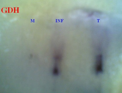
The result of evaluation of GDH enzyme system on PAGE was shown in Figure 1. L. infantum has two intensive bands with Relative Factor (RF) of 0.35 and 0.47 and L. tropica showed a clear band with RF 0.37 and a band with RF 0.5, but there is only a faint band with RF 0.3 in L. major.
There are some smear on PAGE of HK. The HK enzyme system created thick bands using the polyacrylamide matrix, but in the cellulose acetate matrix the bands became sharper and clearer.
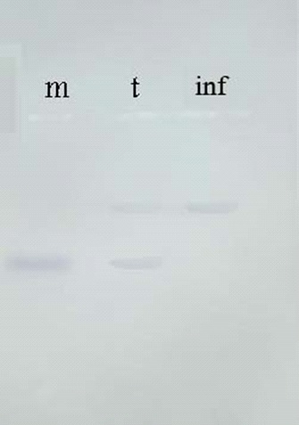
The result of HK enzyme system on cellulose acetate was shown in Figure 2. L. major showed one bands with RF 0.3 and L. tropica showed two bands with RF of 0.18 and 0.3 and I showed one band with RF of 0.18.
Discussion
Leishmaniasis is a tropical disease caused by a variety of parasites from the genus Leishmania. It is transmitted to mammalian hosts by sandflies of the genus Phelebotomus in the Old World and Lutzomia in the New World. Leishmaniasis has a wide specterum of clinical manifestations, including a simple to disseminated skin lesion, and in some species mucocutaneous or visceral symptoms [7,9,11]. The macrophages are the main target cells for Leishmania, but some documents confirmed that fibroblasts may infected with Leishmania [12]. Determining the characteristics of the Leishmania parasite is critical in order to identify vectors and reservoirs of the disease in endemic or epidemic foci. About 30 species of Leishmania are known, of which at least 20 are capable of infecting humans [2]. All of these species are morphologically similar and microscopic examination is only suitable for genus identification and is not able to detect species [10]. Molecular, biochemical and serological tools could be differentiate various species of Leishmania. Among these methods, the method of comparing parasite isoenzymes, which is an accurate method based on the parasite phenotype, is of particular importance. In this method, which is also known as fingerprint method, up to 25 enzyme systems can be studied [4,8,10]. Since this valuable technique is very time consuming and costly, it is necessary to consider the cost-effectiveness of doing it. To achieve this goal, it is necessary to select enzymes that have the lowest cost and the highest power to differentiate between species.
In this study, two systems, GDH and HK, which are active in the metabolic pathways of Leishmania parasite, were studied and in both systems, the power to differentiate between the three most common species in the old world was fully observed. Both systems were able to differentiate L.tropica, L. major and L. infantum.
In some studies the enzymes MDH, NH 1, NH2, PGM, GPI, ME, and 6PGD could differentiate L. major and L. tropica from L. infantum though the enzymes MDH, NH and GPI were found to be more efficient in characterizing these Organisms [5]. The result of HK characterization of Leishmania on cellulose acetate was demonstrated that this method is highly efficient in comparison with PAGE. In contrast, the GDH system in polyacrylamide matrix has a better differentiation ability compared to electrophoresis on cellulose acetate.
Conclusion
Since the isoenzyme electerophoresis technique is very time consuming and costly, it is necessary to consider the cost-effectiveness of doing it. It could be recommended that GDH and HK are suitable systems for differentiate L. major, L. tropica and L. infantum.
Declarations
Author contribution statement
Shirin Memarzadeh, Arghavanvafafar, Khadijehkhanaliha and Farhad Hanjani performed the experiments. Saied Hatam cooperate to edit and analyses the data.
Gholamreza Hatam: Conceived and designed the experiments; performed the experiments; Analyzed and interpreted the data; contributed reagents, materials, analysis tools or data; Wrote the paper.
Acknowledgment: The authors would like to thank the Vice Chancellors of Research of Shiraz University of Medical Sciences, Shiraz, Iran for financial support (Grant No: 19847). This work was extracted from the MD Thesis of Shirin Memarzadeh.
Competing interest statement: The authors declare no conflict of interest.
References
- Ardehali S, Moattari A, Hatam G, Hosseini S, Sharifi I. Characterization of Leishmania isolated in Iran: 1. Serotyping with species specific monoclonal antibodies. Acta tropica. 2000; 75: 301-307.
- Claborn DM. The biology and control of leishmaniasis vectors. Journal of global infectious diseases. 2010; 2: 127.
- Gadisa E, Genetu A, Kuru T, Jirata D, Dagne K, et al. Leishmania (Kinetoplastida): Species typing with isoenzyme and PCR–RFLP from cutaneous leishmaniasis patients in Ethiopia. Experimental parasitology. 2007; 115: 339-343.
- Hatam G, Ardehali S, Riyad M, Bichichi M, Guessous-Idrissi N, et al. Isoenzyme characterization of Iranian Leishmania isolates from cutaneous leishmaniasis. 2005.
- Hatam G, Hosseini S, Ardehali S. Dermotropic isolates of Leishmania infantum in Iran. Transactions of the Royal Society of Tropical Medicine and Hygiene. 1997; 91: 440-440.
- Hatam G, Hosseini S, Ardehali S. Isoenzyme studies in characterization of Leishmania isolated in Iran. Iran J Med Sci. 1999; 24: 8-13.
- Hepburn NC. Cutaneous leishmaniasis: An overview. Journal of postgraduate medicine. 2003; 49: 50.
- Rahi AA, Ali MA, Al-Marjani MF. Characterization of Leishmania species by using Isozyme analysis. Ready PD. (2014). Epidemiology of visceral leishmaniasis. Clinical epidemiology. 2015; 6: 147.
- Reithinger R, Dujardin JC, Louzir H, Pirmez C, Alexander B, Brooker S. Cutaneous leishmaniasis. The Lancet infectious disease. 2007; 7: 581-596.
- Rioux J, Lanotte G, Serres E, Pratlong F, Bastien P, Perieres J, et al. Taxonomy of Leishmania. Use of isoenzymes. Suggestions for a new classification. Annales de parasitologie humaine et comparee. 1990; 65: 111-125.
- Shirian S, Oryan A, Hatam GR, Daneshbod Y. Mixed mucosal leishmaniasis infection caused by Leishmania tropica and Leishmania major. Journal of Clinical Microbiology. 2012; 50: 3805- 3808.
- Yektaeian N, Mehrabani D, Sepaskhah M, Zare S, Jamhiri I, et al. Lipophilic tracer Dil and fluorescence labeling of acridine orange used for Leishmania major tracing in the fibroblast cells. Heliyon. 2019; 5: e03073.