
Journal of Clinical Images and Medical Case Reports
ISSN 2766-7820
Short Report - Open Access, Volume 3
Bilateral giant cervical cystic lymphangioma in an infant
Charu Tiwari1; Nitin kumar Borkar2*; Rohit Meshram3
1 Assistant Professor, Department of Paediatric Surgery, All India Institute of Medical Sciences, Raipur, CG, India.
2 Additional Professor and Head, Department of Paediatric Surgery, All India Institute of Medical Sciences, Raipur, CG, India.
3 Senior Resident, Department of Paediatric Surgery, All India Institute of Medical Sciences, Raipur, CG, India.
*Corresponding Author: Nitinkumar Borkar
Additional Professor and Head, Department of
Paediatric Surgery, All India Institute of Medical
Sciences, Raipur, CG, India.
Email: drnitinborkar25@gmail.com
Received : Jan 03, 2022
Accepted : Feb 17, 2022
Published : Feb 24, 2022
Archived : www.jcimcr.org
Copyright : © Borkar N (2022).
Abstract
Cystic Hygroma is a rare congenital lymphatic malformation; more so rare is the bilateral cystic Hygroma. Bilateral Giant cystic Hygroma is a potential life-threatening congenital malformation as it may cause airway obstruction, difficulties in nursing and feeding and failure to thrive. We present a case of a female infant with bilateral giant cystic hygroma.
Keywords: cystic hygroma; bilateral; giant.
Citation: Tiwari C, Borkar N, Meshram R. Bilateral giant cervical cystic lymphangioma in an infant. J Clin Images Med Case Rep. 2022; 3(2): 1690.
Introduction
Cystic hygroma, also known as lymphangioma, is a rare congenital malformation of the lymphatic system, most commonly seen in head and neck region [1]. Although lymphangiomas are benign lesions, they can compress the vital structures in the neck or can cause complications like intracystic hemorrhages, cyst rupture or infections. Giant cystic hygroma of neck and oral cavity may present with respiratory distress and dysphagia [2]. We present a case of a female infant with bilateral giant cystic hygroma.
Case summary
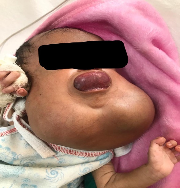
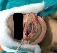
A 2 months female child was admitted with bilateral giant cystic cervical swelling, tachypnea, failure to thrive and fever (Figure 1). Tongue was protruded and cystic swelling could be seen at floor of the mouth. Clinical diagnosis of bilateral giant cervical cystic Hygroma (lymphovascular malformation) was made. Patient was started on nasogastric feeds and intravenous antibiotics. Local Ultrasonography revealed multicystic lymphatic cysts in bilateral neck and floor of mouth. Computed Tomography of neck and upper thorax confirmed the diagnosis and ruled out extension into the thoracic cavity. Patient was taken for surgical excision after stabilization. Intubation was difficult and was possible only after 50 cc of lymphatic fluid was aspirated from the floor of the mouth. Complete excision of the Lymphangioma was done form bilateral cervical region and also from floor of the mouth (Figure 2). The patient was kept on elective mechanical ventilation for 3 days post-surgery and then extubated. Post-operative recovery was uneventful and she was discharged on 15th post-operative day. There is no recurrence at 6 months of follow-up.
Discussion
The problems with bilateral giant cervical lymphangiomas in neonates are usually related to airway obstruction, feeding issues, disfigurement and difficult intubation. In recent times, with better antenatal screening, such cases are diagnosed antenatally and are delivered in institutions with appropriate perinatal care facilities. The usual primary management is intralesional Sclerotherapy for large lesions in neonates and infants and/or wait and watch for smaller lesions.
This case was unique that there was no antenatal diagnosis; delayed presentation at two months; significant disfigurement was present with tongue protrusion, inadequate feeding resulting in failure to thrive and presentation with mild tachypnea. Airway was also compressed making the intubation difficult. The difficult intubation was taken care of by aspirating fluid from the floor of the mouth, thereby opening the airway enough for intubation. As there was no intrathoracic extension, complete excision was possible. We proceeded directly to surgical excision as the lesion was large and bilateral causing difficulty in nursing and feeding by the parents and disfigurement of the baby.
References
- Guruprasad Y, Chauhan DS. Cervical cystic hygroma. Journal of Maxillofacial and Oral Surgery. 2012; 11: 333-336.
- Karakas O, Karakas E, Boyaci FN, Murat Yildizhana, Songul Demir, et al. Cervicomediastinal Giant Cystic Hygroma. A Case Report. J Clin Med Res. 2013; 5: 61-63.