
Journal of Clinical Images and Medical Case Reports
ISSN 2766-7820
Case Report - Open Access, Volume 3
Sporadic Burkitt’s lymphoma presenting as intussusceptions in a 12-year-old Chinese boy
Bingsong Wang1; Benhong Xiang2; Ming Cao3; Kaihu Yao4; Qinghong Meng4*
1 Department of Pediatrics, Wuhu No.1 People’s Hospital, Anhui, China.
2 Department of Pediatric Surgery, Wuhu No.1 People’s Hospital, Anhui, China.
3 Department of Pathology, Wuhu No.1 People’s Hospital, Anhui, China.
4 Beijing Pediatric Research Institute, Beijing Children’s Hospital, Capital Medical University, National Center for Children’s Health, Beijing, China.
*Corresponding Author: Qinghong Meng
Beijing Children’s Hospital, Capital Medical University, National Center for Children’s Health, Beijing,
China.
Email: mengqinghong@aliyun.com
Received : Jan 29, 2022
Accepted : Feb 22, 2022
Published : Mar 01, 2022
Archived : www.jcimcr.org
Copyright : © Meng Q (2022).
Abstract
Intussusception is usually seen at infancy and early childhood, and majority of cases occur in children younger than 2 years. The cause of intussusceptions is typically unknown in children. We report a case of a 12-year-old boy who presented with a 1-month history of intermittent abdominal pain and was found to have an intussusceptions causing by sporadic Burkitt’s lymphoma (BL). This case demonstrates the importance of considering BL as the lead point for chronic intussusceptions, though rare, in children of an atypical age group.
Citation: Wang B, Xiang B, Cao M, Yao K, Meng Q. Sporadic Burkitt’s lymphoma presenting as intussusceptions in a 12-yearold Chinese boy. J Clin Images Med Case Rep. 2022; 3(3): 1702.
Background
Intussusception is a condition in which one segment of bowel telescopes into an adjacent segment [1]. Intussusception is more common among children than adults, in children it is one of the most common acute abdomen [2]. Intussusception is usually seen at five months of life, peaks at four to nine months, and then gradually declines at around 18 months [1]. Majority of cases occur in children younger than 2 years.
The causes of child intussusception are always unclear. About 75% of cases of intussusception in children arise from an unknown cause. Underlying pathological causes of intussusception can be identified in only 25% of cases [3]. These include Meckel’s diverticulum, polyps, duplications, mesentery cysts, intestinal hematoma and lymphoma [4]. We report a case of a 12-year-old boy who presented with a 1-month history of intermittent abdominal pain and was found to have an intussusceptions causing by sporadic Burkitt’s lymphoma (BL). This case demonstrates BL as the lead point for chronic intussusceptions, though rare, in children of an atypical age group in regions outside Africa.
Case presentation
A 12-year-old boy presented to our hospital with a 1-month history of intermittent abdominal pain below the xiphoid process and around umbilicus. Pain had been present for 1 year and was mild. For 4 days before admission, he had an exacerbated colicky abdominal pain and hematochezia. On physical examination we found his abdomen was soft with a left lower abdominal mass, measuring 6 X 4 cm. His vital signs were stable. Plain abdominal X-ray radiograph shows multiple air-fluid levels (Figure 1). Ultrasound and computed tomography (Figure 2) of the abdomen showed dilated loops of small bowel and a target-sign, suggestive of small bowel intussusceptions.


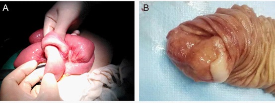
During an explorative laparotomy, a 20 cm enteric intussusception was found (Figure 3A) and manually reduced. After reduction, a 4.2 X 3 X 2.6 cm mass was uncovered at the beginning of the jejunum, then all the affected segments was resected (Figure 3B). Histologic analysis of the resected mass showed the medium to large-sized lymphoid cells admixed with scattered tingible body macrophages imparting a “starry sky” appearance (Figure 4A-D). Immunostaining for CD20, CD10, LCA, CD79α were positive. The Ki-67 labeling index exceeded 90%. Sporadic Burkitt’s lymphoma (BL) presenting as intussusceptions was the final diagnosis. The patient recovered uneventfully after surgery and received chemotherapy. At follow-up 4 year later, he had no recurrence.

Discussion
Intussusception is usually seen at young infants. Chronic intussusceptions as a clinical entity is poorly recognized and rarely included in the differential diagnosis of prolonged abdominal symptoms for older child [5]. This case demonstrates the importance of considering chronic intussusception, though rare, in children of an atypical age group.
The cause of intussusceptions is typically unknown in children, while in adults a pathological lead point due to malignant tumors is often present. BL is categorized into endemic, sporadic, and immunodeficiency-associated subtypes [6]. Endemic BL is the most common childhood cancer in tropical Africa [7]. Sporadic BL attacks regions outside Africa, and majority of cases occur in the United States (US) and Western Europe. Sporadic BL frequently involves distal ileum, caecum and mesentery, presenting symptoms include abdominal swelling due or pelvic mass, abdominal pain and intussusception-related symptoms of bowel obstruction [8]. Intussusception in itself is a very rare presentation of BL. To our knowledge, this is the first reported case of child intussusception due to BL in China. The case was unusual in that the child did not present with the typical clinical features of BL, and he was not a high-risk demographic for this rare disease. Early diagnosis and treatment make for a good prognosis.
References
- Jain S, Haydel MJ. Child Intussusception. [Updated 2021 Jul 17]. In: StatPearls [Internet]. Treasure Island (FL): StatPearls Publishing. 2022.
- Byrne AT, Geoghegan T, Govender P, Lyburn ID, Colhoun E, Torreggiani WC. The imaging of intussusception. Clin Radiol. 2005; 60(1): 39-46.
- Ng JY, Thompson RJ, Lam A, Nigam S. Sporadic Burkitt’s lymphoma masquerading as an intussuscepted Meckel’s diverticulum in a 9-year-old child. BMJ Case Rep. 2018: bcr2018224333.
- Sorantin E, Lindbichler F. Management of intussusception. Eur Radiol. 2004; 14 Suppl 4: L146-54.
- Saad AAM, Kkalid T, Abbas M, Salih KMA. Rare presentation of chronic ileocecal intussusception secondary to Burkitt’s lymphoma in three years Sudanese boy: a case report and literature review. Pan Afr Med J 2018; 31: 57.
- Crombie J, LaCasce A. The treatment of Burkitt lymphoma in adults. Blood. 2021; 137(6): 743-750.
- Quintana MDP, Smith-Togobo C, Moormann A, Hviid L. Endemic Burkitt lymphoma - an aggressive childhood cancer linked to Plasmodium falciparum exposure, but not to exposure to other malaria parasites. APMIS. 2020; 128(2): 129-135.
- Simson R, Planner A, Alexander R. Adult Burkitt’s lymphoma presenting as intussusception: first UK case report. Ann R Coll Surg Engl. 2017; 99(7): e206-208.