
Journal of Clinical Images and Medical Case Reports
ISSN 2766-7820
Clinical Image - Open Access, Volume 3
Right ventricular myxoma: An incidental finding on CT chest lung screen
Rebecca DeBoer, DO1*; Christopher Reggio, DO1; Charnjeet Sandhu, MD2; Julian Diaz Fraga, MD2
1 Department of Medicine, Reading Hospital, Reading, PA 19611, USA.
2 Department of Cardiology, Reading Hospital, Reading, PA 19611, USA.
*Corresponding Author: Rebecca DeBoer
Department of Medicine, Reading Hospital, Reading, PA 19611, USA.
Email: Rebecca.deboer@towerhealth.org
Received : Jan 20, 2022
Accepted : Mar 02, 2022
Published : Mar 09, 2022
Archived : www.jcimcr.org
Copyright : © DeBoer R (2022).
Citation: DeBoer R, Reggio C, Sandhu C, Fraga JD. Right ventricular myxoma: An incidental finding on CT chest lung screen. J Clin Images Med Case Rep. 2022; 3(3): 1725.
Description
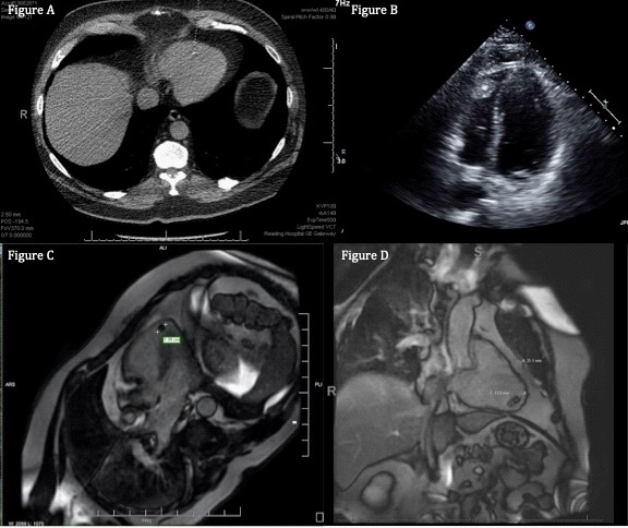
With the rise in screening modalities, incidental findings increase. We present a case of a computed tomography (CT) chest lung cancer screen that resulted in the diagnosis of a right ventricular (RV) myxoma. Echocardiogram and cardiac magnetic resonance imaging (CMR) led to this diagnosis.
A 57-year-old Caucasian male with a 60-pack year history presented to his primary care provider for an annual exam. CT chest revealed a faint calcification in the RV apex (Figure 1A). In subsequent transthoracic echocardiogram, attached to the RV apex free wall was a 2.4 cm X 1.5 cm heterogeneous mass with central calcification suggestive of RV myxoma (Figure 1B). Cardiac magnetic resonance imaging (CMR) showed minimal rim enhancement of a 2.1 X 2.5 X 1.3 cm centrally calcified mass arising from the free wall of the RV apex consistent with myxoma (Figure 1C and 1D). The patient remained asymptomatic with a normal cardiac exam.
Cardiac tumors remain rare [1]. They can be divided into primary or secondary [1]. Most primary tumors are myxomas found in the atria [2]. Only a small portion originates in the right ventricle [2]. Estimated to be 8% in one review [2]. Most patients will be symptomatic [2]. One study found only 13% of patients were asymptomatic [2].
Echocardiography has long been utilized and cardiac magnetic resonance imaging (CMR) has become a more optimal form of imaging cardiac tumors [3]. Due to the improved resolution of CMR, this imaging modality can lead to definitive diagnoses of cardiac masses [4]. CMR can reliably indicate the extent of tumor and attachment to the cardiac wall [4]. CMR can also characterize the anatomy and tissue features, which can differentiate from other masses such as malignant tumors and cardiac thrombi [3].
References
- Braunwald E, Zipes DP, Libby P. Heart disease. Philadelphia, PA: Saunders. 2019: 1866-1877.
- Acebo E, Val-Bernal JF, Gómez-Román JJ, Revuelta JM. Clinicopathologic study and DNA analysis of 37 cardiac myxomas: a 28-year experience. Chest. 2003; 123(5): 1379-85. doi: 10.1378/chest.123.5.1379. PMID: 12740251.
- O’Donnell DH, Abbara S, Chaithiraphan V, Yared K, Killeen RP, Cury RC, et al. Cardiac tumors: optimal cardiac MR sequences and spectrum of imaging appearances. AJR Am J Roentgenol. 2009; 193(2): 377-87. doi: 10.2214/AJR.08.1895. PMID: 19620434.
- Gulati G, Sharma S, Kothari SS, Juneja R, Saxena A, Talwar KK. Comparison of echo and MRI in the imaging evaluation of intracardiac masses. Cardiovasc Intervent Radiol. 2004; 27(5): 459-69. doi: 10.1007/s00270-004-0123-4. Epub 2004 Jun 16. PMID: 15383848.