
Journal of Clinical Images and Medical Case Reports
ISSN 2766-7820
Research Article - Open Access, Volume 3
Remodelled multiple linear regression equation for stent size calculations of left anterior descending artery (LAD-proximal) dimensions with single vessel disease
Divia Paul A1*; Ashraf SM2; Ranajit Das3; Ashwini Prabhu3
1 Department of Anatomy, Yenepoya Medical College, Karnataka, India.
2 Department of Cardio Vascular Sciences, Sahakarana Hrudayalaya, Pariyaram Medical college, Kerala, India.
3 Yenepoya Research Centre, Yenepoya University, Deralakatte, Mangalore -575018, Karnataka, India.
*Corresponding Author: Divia Paul A
Assistant Professor, Department of Anatomy, Yenepoya Medical College, Yenepoya (Deemed to be
University), University Road, Deralakatte, Mangalore, Karnataka 575018, India.
Email: divia_manoj@yahoo.com
Received : Feb 09, 2022
Accepted : Mar 08, 2022
Published : Mar 15, 2022
Archived : www.jcimcr.org
Copyright : © Paul AD (2022).
Abstract
Purpose of research: To validate the Multiple Linear Regression Equation (MLR) of predicted dimensions of left anterior descending coronary artery (LAD-o, p) and left circumflex artery (LCx-o, p) by remodelling the equation based on the percentage of stenosis present in the artery segments. This helped us to go forward with an objective to explore the possibility of explaining the relationship with a MLR formula, to estimate the reference diameter of a stenosed coronary artery when the other two arteries are normal.
Materials and methods: Patients who undergo Percutaneous Coronary Angioplasty (PCI) procedure were included from single study centre of South India after obtaining the ethical clearance through proper channels. Total n=104 cases who have undergone PCI in LAD-p were studied prospectively. Informed consent was obtained. Exclusion criteria’s were post CABG and post PCI patients. The parameters assessed and categorized were coronary artery measurements of non-diseased segments from Single Vessel Diseased Coronaries (SVD) after excluding the diseased segments.
Results and conclusions: The initial participant predicted equation for LAD-p was remodelled after validation by considering the difference between stent size estimated (calculated) and stent reduction category. Participant predicted equation: LAD-p=1.092+0.302*LMCA+0.379 * LCx-p. F (2,101) = 1340, p<0.0001; with an R2 of 0.9637. Age and gender had no significant change while taking in to consideration for remodelling (p =0.17).
PCI was found to be safe and effective in our routine clinical setting during the validation of the estimated MLR formula and was valuable in detecting the artery diameter in stenosed SVD of LAD-p.
Keywords: Percutaneous coronary angioplasty; Multiple linear regression equation; Left anterior descending artery; Single vessel diseased coronaries; Stenting strategies.
Citation: Paul AD, Ashraf SM, Das R, Prabhu A. Remodelled multiple linear regression equation for stent size calculations of left anterior descending artery (LAD-proximal) dimensions with single vessel disease. J Clin Images Med Case Rep. 2022; 3(3): 1735.
Introduction
Coronary Artery Disease (CAD) is the leading cause of death worldwide. CAD is one of the most common causes of morbidity and mortality especially in the developing countries, which accounts for more than one-third of the total deaths. Coronary artery diameter is one of the most important factors that affect the procedure and outcome of Percutaneous Coronary Angioplasty (PCI) and Coronary Bypass Operations (CABG) [1]. Noninvasive Quantitative Coronary Angiography (QCA) has become an important imaging tool in the evaluation of patients with and at risk of CAD [2,3]. Invasive coronary angiography is the gold standard for establishing the presence, location and severity of CAD [4,5]. Left Main Coronary Artery (LMCA) length can be variable, and shorter in comparison with other main coronary arteries. So, atherosclerotic plaques can occupy the whole vessel in some cases. In such cases, it is difficult to determine the degree of narrowing caused by the plaque as possible to reference artery diameter determination is difficult. Similarly, confirmation of coronary artery dimensions will be troublesome if a plaque builds up as a long segment in the Left Anterior Descending Artery (LAD) or Left Circumflex Artery (LCx) starting from the ostium. Cerain studies attempted to elucidate the correlation between the diameters of coronary arteries at bifurcation levels using Murray’s law or Finet’s formula [6,7]. In this study, we aimed to validate the multiple linear regression equation of predicted dimensions of ostial and proximal segments of left anterior descending coronary artery (LAD-o,p) and left circumflex artery (LCx-o,p) and to remodel the predicted equation based on the percentage of stenosis present in the artery segments by a single study centre approach .This helped us to go forward with an objective to explore the possibility of explaining the relationship with a multiple linear regression formula, which may then be used to estimate the reference diameter of a stenosed coronary artery when the other two arteries are normal without stenosis.
Materials and methods
A cross-sectional study was conducted at single centre of South India. Hospital was purposely selected according to the number of cardiac patients identified by them. Patients who undergo Percutaneous Coronary Angioplasty (PCI) procedure owing to abnormalities detected during the QCA were included after obtaining their informed consent. All ethical principles for human research were followed, and ethical approval was obtained from the institutional ethics committee of the parent institute (YUEC/343/2016 Date-01.10.2016) and of the hospital from where data was collected (No.G1.2747/12/ACME Date22.08.2017). By participating in this research, patients were informed that they are doing social contribution and will not get any direct benefits. The study may result in better treatment for the disease. The patients were informed that the knowledge that we get from doing this research may be published or presented in scientific forums in order that other interested people may learn from our research. Confidential information will not be shared. Informed consents were obtained.
Total n=104 cases who have undergone the evaluation for CAD followed by PCI in LAD-p (proximal segment) were included by convenience sampling technique. Though we attempted to validate all the MLR equations, calculated from N=625 samples of SVD cases from a total of QCA images of 3855 cases who have undergone the evaluation for CAD by convenience sampling technique by an initial study at four centres of South India. Sufficient samples with defined endpoints were only available for LAD-p for the present study. So remodelling was only possible for the equation for LAD-p (n=104 samples). Twenty seven cases were excluded by the authors for the following reasons: presence of atheromatous changes, presence of coronary artery origin abnormalities, absence of the LMCA, double vessel disease and altered coronary anatomy due to operation which leaded to difficulty in evaluation. Thus, 104 cases made up the study population (77 men and 27 women; age range, 30–75 years). The age of the study subjects was given a cut-off at 75 years owing to marginal benefits marked during the follow-ups. Hence, a conservative approach is proven to be appropriate for the aforementioned age, which itself indicates a poor prognosis with an average yearly mortality rate of 33–35% [8].
Data collection
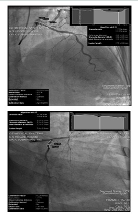
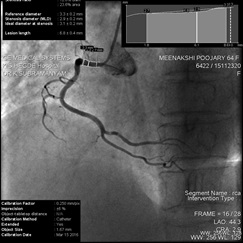
Procedures: The medical records of all eligible study patients were reviewed, and baseline characteristics such as age, sex, medical history, concomitant medications, and details of clinical presentation, vital signs, details of electrocardiogram and echocardiogram, and laboratory assessments were collected. Pre-procedural QCA was performed in all cases according to validated standards. Pre-procedural parameters that were recorded included: (a) number of target vessels and lesions involved; (b) target vessel details; (c) ostial involvement of LMCA; (d) presence of thrombus, chronic total occlusion, or bifurcation lesions in the segments; (e) degree of calcification (none, mild, moderate, and severe); (f) complexity of lesions according to the American College of Cardiology (ACC)/American Heart Association (AHA) [9]; and (h) diameter stenosis. The PCI procedure was performed with standard techniques and catheters using a femoral approach in 90 cases and Radial approach in 14 cases. Stenting strategy, lesion preparation, and the number of stents used were left to concerned operator’s discretion. The number of stents used, details of length and diameter of the stents, and stent overlap were recorded.
All images were digitally stored and deidentified. Post dilatation was at the discretion of the operator and was performed mostly using non-compliant balloons. Dual antiplatelet therapy and other medications per the ACC/AHA guidelines were administered. The LMCA diameter was taken at the midpoint between the ostium and the bifurcation level into the LAD and LCx by using catheter calibrations. The maximum diameter region was taken for assessment. However, in arteries with minimum calibre, measurements were taken at or near the ostium. The diameter calibrations of the LAD and LCx were taken at the ostium and the proximal segment of the LAD by using catheter calibrations. The maximum diameter region was taken for assessment. All QCA images were re-reviewed by two cardiologists from the centre for the definition of other two normal vessels among SVD cases and for the subsequent quantitative analysis by the double blinding method. Both the observers were blinded regarding the patient identity, and interobserver variability was accounted during remodelling of the equation.
Multiple linear regression equation was formulated from the initial study by the authors for ostial and proximal segments of LAD and LCx and we observed a strong correlation between LMCA dimensions and LAD and LCx dimensions (r = 0.526**, p < 0.001* and r= 0.469**, p < 0.001*, respectively) [10]. The equations were as follows which was calculated from N=625 samples of SVD cases from a total of QCA images of 3855 cases who have undergone the evaluation for CAD by convenience sampling technique from the initial study for 2 years.
• Participant predicted equation: LAD-o=1.110+0.335*LMCA+0.357*LCx-o [F (16.184), p<0.001; with an R2 of 0.049 respectively] • Participant predicted equation: LAD-p =1.337+0.292*LMCA+0.395*LCx-p [F (19.205), p<0.001; with an R2 of 0 .058 respectively] • Participant predicted equation: LCx-o= 1.158+0.415*LMCA+0.120*LAD-o [F (38.694), p<0.001; with an R2 of 0.110 respectively] • Participant predicted equation: LCx-p= 1.204+0.388*LMCA+0.137*LAD-p [F (42.110), p<0.001; with an R2 of 0.119 respectively].
Among all predictions, both of the predictor variables are significant with p < 0.05, which indicates both the variables are significantly contributing to the dependant variable. For validation, after predicting the dimensions of the respective diseased segment of SVD artery, the percentage of stenosis was noted down and graded respectively as the following ranges based on the statistical inferences.
1. Grade A: Less than 40% stenosis with no change in calculated stent size 2. Grade B: 41-60% stenosis with 0.1 – 0.2 mm difference from the calculated stent size 3. Grade C: 61-80% stenosis with 0.2 – 0.3 mm difference from the calculated stent size 4. Grade D: 81% to complete total occlusion with retrograde filling of coronary artery with 0.3 – 0.4 mm difference from the calculated stent size.
After grading the artery, according to the percentage of stenosis present in the segment, the readjusting of the equation according to the respective grades using the appropriate statistical methods of scoring. Age factor was also considered for remodelling by giving appropriate territory fraction level importance to age. Remodelled equation was again tested to validate the suitability of application among each grade. The size of stent used for the patient was noted down to see the accuracy by comparing with predicted dimension from the equation. The accuracy indicated the external validity of the equation which can be generalisable to the target population. Among 104 patients, 86 patients had followed the regular clinic visit follow-up at 6 months and remaining patients, 18 had telephonic follow up.
Statistical analysis
Statistical analysis was conducted using Graph Pad Prism v9. Association between LMCA, LAD, LCx diameters, age and gender was assessed in a multiple regression framework. The age and gender were compared using t-test. Multiple linear regression analysis was further used to develop a model to elucidate the relationship between LMCA, LAD and LCx diameters. The model fit was assessed using appropriate residual and goodness-of-fit statistics.
Results
The mean age of the patients was 55.50 ± 6.49 years. Physical and demographic parameters were assessed. The average weight was 65.20 ± 3.09 kg (range, 90.00–37.00 kg) and height was 170.15 ± 4.27 cm (range, 190.00–135.00 cm). We couldn’t validate the equation for the SVD cases of LAD-o, LCx-(o,p) due to unavailability of the sufficient cases to validate a MLR equation which was fitting to our inclusion criteria during the stipulated study period of one year
Regression analysis and prediction
Age (t=1.44, p=0.15) and gender (t=1.39, p=0.17) had no significant association with SVD parameters and so were not considered during remodelling. The participant predicted equation for LAD-p (LAD-p=1.337+0.292*LMCA+0.395*LCx-p) was remodelled after validation by subtracting stent reduction category (B) from the stent size estimated (calculated) (A). We surmised that the difference (C) between A and B should be close to stent used (D). Employing this strategy, we could reduce the Coefficient of Variation (CV) in stent used (D) to 1.068. We then employed the difference (C) in a multiple regression framework alongside LMCA and LCx-p to statistically determine the new intercept and slopes b1 and b2 for LMCA and LCx-p respectively. The new intercept was 1.092 and the slopes for LMCA and LCx-p were 0.302 and 0.379 respectively. Based on this the new participant predicted equation was: LADp=1.092+0.302*LMCA+0.379*LCx-p. The multiple regression coefficient (R2 ) for the new equation was 0.9637, very similar to the original equation (R2 =0.9936). This model was validated using Least squares method (F-value (2,101) =1340 (p<0.0001). Sum of squares (SS) of the residuals for this model, however, was discernibly higher (0.533) than the original model (0.093), which can be visible in the residual plots (Figure 1 and Figure 2). For every unit increase in the LMCA, we expect a 0.302 mm increase in LAD-p, holding all other variables constant, and for every unit increase in LAD-p, we expect a 0.379 mm increase in LCx-p, holding all other variables constant (p < 0.001; with an R2 of 0.9637).
Discussion
Knowing the dimensions of a coronary artery is crucial for appropriate stent sizing. Inappropriate measurements can predispose the patients to a higher rate of post-PCI complications such as in-stent restenosis within 6 months of the procedure. Increased mortality in women after revascularization has been attributed to gender-related differences in the coronary size [1,11] and the artery size can be related to the outcomes after PCI [12,13]. The positive correlation [10] indicated that a regression analysis can be carried out by incorporating the measurements. Geometry of the ideal bifurcation is described by Murray’s law, which states that the cube of the radius of a parent vessel equals the sum of the cubes of the radii of the daughters.
Both studies using this law at the LMCA branching level reported that it was successful in defining coronary artery diameters [14,15].
In right-dominant circulation, there was a linear correlation between the diameter of the right coronary artery (RCA) and its right Posterior Descending (PD) and Posterior Left Ventricular (PLV) branches. Also, there was a positive correlation between the size of the LMCA with LAD, LCx along with the left Posterior Descending Artery (PD) among left-dominant circulation [16]. A strong positive correlation was observed between the diameter of the LMCA with prox LAD and LCx. Pearson’s correlation coefficient of LAD was 0.623**(mm) and LCx was 0.428**(mm) respectively [17]. A strong correlation between the cross-sectional areas of LMCA with LAD and LCx was assessed (r = 0.779, p < 0.001 and r = 0.678, P < 0.001, respectively) at the bifurcation level, and a regression equation was formulated to predict LMCA dimensions (LMCA= 3.870 + 0.718 × LAD + 0.434 × LCx) [18]. But the present study reveals that the equation cannot be unique for the LMCA as the cases for isolated left main lesions are limited and risk levels are more in those cases. The LMCA and its branches need to be assessed throughly and the equations cannot be gender specific.
In our study, we found the dimensions of the LMCA, LAD and LCx, which can have a wide clinical perspective. The determinants of coronary vessel dimensions were total or regional left ventricular mass, age and body surface area by multivariate regression analysis. Total left ventricular mass was used for Total Coronary Cross-Sectional Area (TCSA) because the percent territory score corresponding to TCSA was 100% [19]. Multiple regression equations were formulated with mean diameters of LMCA, prox LAD and prox LCx. A territory fraction was added for age of the patient. The volume of barium sulphate-gelatin injected into postmortem coronary arteries was measured and there was a significant linear relationship between the sums of the cross-sectional areas of the major coronary arteries (prox LAD and proxLCx) and the capacities of the coronary arterial tree [20]. Studies investigating cross-sectional areas of coronary arteries were mainly carried out with the purpose of investigating the LMCA. The distal LMCA sectional area was reported as 14 ± 5 mm2 in an Multiple-Detector Computed Tomography (MDCT) study [21]. However, the LMCA cross-sectional area to be 12.4 ± 4.4 mm2 by few authors [22], which is smaller than that of the study by Zeina et al [21]. The present study suggests it is better to take coronary artery measurements in diameter calibrations than the cross-sectional area as the former can help to calculate the dimensions of the coronaries during the PCI procedures more easily. This in turn can help to assign the stent size in a diseased artery according to the grade of stenosis. Age factor was not added as a territory fraction in the present study because there was no correlation found between coronary artery measurements and the patient’s age. Similar findings were reported in studies by Hutchins et al [23] and Zeina et al. [21] Some other studies found a slight increase in coronary arteries with age [24,25] and also age-related decrease in vasodilatation of the arteries [19]. However, the sample sizes were small in these studies to draw appropriate conclusions; the present study had a larger sample size which proved the accuracy and relevance about age related conclusions.
Conclusion
In conclusion, PCI was found to be safe and effective in our routine clinical setting during the validation of the estimated MLR formula of the present study and was valuable in detecting the artery diameter in stenosed LAD and can avoid major stent malapposition, tissue prolapse, and edge dissections.
Limitations
We couldn’t do an OCT guided PCI which could have provided us with better clinical outcomes. Randomized studies comparing OCT-guided PCI versus angiography are warranted to further establish the efficacy and safety of the MLR equation established in Indian settings.
Summary
• Remodelled Multiple Linear Regression Equation (MLR) of Left Anterior Descending coronary artery (LAD-proximal) based on the percentage of stenosis present in the artery segments • The findings will help to valuable in detecting the artery diameter in single vessel diseased (SVD) cases of LADproximal when Left Main Coronary (LMCA) and left circumflex arteries are disease free segments • This has important clinical relevance as it helps for improved procedural outcomes. • This can be also an add-up to the global data pool on the matter.
Declarations
Conflict of interest: All authors have none to declare. We declare there is no conflict of interest and no financial supports or grants were received for conduction of the study
Funding: This research received no specific grant from any funding agency in the public, commercial, or not-for-profit sectors.
Author contributions: All authors hereby declare that their contribution was equal towards the formation of the manuscript.
Acknowledgements: All authors appreciate the great effort of chief cardiac technicians of the cardiac catheterization laboratory of, Pariyaram Medical College, Kannur, Kerala, India; for their timely help and assistance in the conduct of this study. All authors appreciate the great effort of Mr. Jessil Jose, FIA (FELLOW OF INSTITUTE OF ACTUARIES), Head of Reserving company: Advent Underwriting Limited, United Kingdom and Dr. Ranajith Das,Yenepoya Research Centre, Mangalore, Karnataka, India for analysing and verifying the data of this study
References
- O’Connor NJ, Morton JR, Birkmeyer JD, Olmstead EM, O’Connor GT. Effect of coronary artery diameter in patients undergoing coronary bypass surgery. Circulation. 1996; 93: 652-655.
- Kohsaka S, Makaryus AN. Coronary angiography using noninvasive imaging techniques of cardiac CT and MRI. Curr. Cardiol. Rev 2008; 4: 323-330.
- Kosar P, Ergun E, Öztürk C, Kosar U. Anatomic variations and anomalies of the coronary arteries: 64-slice CT angiographic appearance. Diagn Interv Radiol. 2009; 15: 275.
- Levin DC. Invasive evaluation (coronary arteriography) of the coronary artery disease patient: Clinical, economic and social issues. Circulation. 1982; 66: III71-9.
- Ryan TJ. The coronary angiogram and its seminal contributions to cardiovascular medicine over five decades. Circulation. 2002; 106: 752-756.
- Saikrishna C, Talwar S, Gulati G, Kumar AS. Normal coronary artery dimensions in Indians. Indian J. Thorac. Cardiovasc. Surg. 2006; 22:159-164.
- Yang F, Minutello RM, Bhagan S, Sharma A, Wong SC. The impact of gender on vessel size in patients with angiographically normal coronary arteries. J. Interv. Cardiol. 2006; 19: 340-344.
- Azad N, Lemay G. Management of chronic heart failure in the older population. Journal of geriatric cardiology: JGC. 2014; 11: 329.
- Krone RJ, Shaw RE, Klein LW, Block PC, et al. Evaluation of the American College of Cardiology/American Heart Association and the Society for Coronary Angiography and Interventions lesion classification system in the current “stent era” of coronary interventions (from the ACC-National Cardiovascular Data Registry). AJC. 2003; 92: 389-394.
- Paul AD, Ashraf SM, Ezhilan J, Vijayakumar S, Kapadiya A, et al. A milestone in prediction of the coronary artery dimensions from the multiple linear regression equation. IHJ. 2019; 71: 328-333.
- Peterson ED, Lansky AJ, Kramer J, Anstrom K, Lanzilotta MJ, National Cardiovascular Network Clinical Investigators. Effect of gender on the outcomes of contemporary percutaneous coronary intervention. AJC. 2001; 88: 359-364.
- Bell MR, Grill DE, Garratt KN, Berger PB, Gersh BJ, Holmes Jr DR. Long-term outcome of women compared with men after successful coronary angioplasty. Circulation. 1995; 91: 2876-2881.
- Saucedo JF, Popma JJ, Kennard ED, Talley JD, Lansky A, et al. Relation of coronary artery size to one-year clinical events after new device angioplasty of native coronary arteries (a New Approach to Coronary Intervention [NACI] Registry Report). AJC. 2000; 85: 166-171.
- van der Waal EC, Mintz GS, Garcia Garcia HM, Bui AB, Pehlivanova M, Girasis C, et al. Intravascular ultrasound and 3D angle measurements of coronary bifurcations. Catheter. Cardiovasc. Interv. 2009; 73: 910-916.
- Zhou Y, Kassab GS, Molloi S. On the design of the coronary arterial tree: A generalization of Murray’s law. Phys. Med. Biol. 1999; 44: 2929.
- Saikrishna C, Talwar S, Gulati G, Kumar AS. Normal coronary artery dimensions in Indians. Indian J. Thorac. Cardiovasc. Surg. 2006; 22: 159-164.
- Shukri IG, Hawas JM, Karim SH, Ali IK. Angiographic study of the normal coronary artery in patients attending ulaimanicenter for heart diseases. Eur Sci J. 2014; 10.
- Verim S, Öztürk E, Küçük U, Kara K, Sağlam M, et al. Cross-sectional area measurement of the coronary arteries using CT angiography at the level of the bifurcation: is there a relationship?. Diagn Interv Radiol. 2015; 21: 454.
- Leung WH, Stadius ML, Alderman EL. Determinants of normal coronary artery dimensions in humans. Circulation. 1991; 84: 2294-2306.
- Rodriguez FL, Robbins SL, Connolly OF. Capacity of human coronary arteries: A postmortem study. Circulation. 1959; 19: 570- 578.
- Zeina AR, Rosenschein U, Barmeir E. Dimensions and anatomic variations of left main coronary artery in normal population: multidetector computed tomography assessment. CAD. 2007; 18: 477-482.
- Christensen KN, Harris SR, Froemming AT, Brinjikji W, Araoz P, Asirvatham SJ, et al. Anatomic assessment of the bifurcation of the left main coronary artery using multidetector computed tomography. SARA. 2010; 32: 903-909.
- Hutchins GM, Bulkley BH, Miner MM, Boitnott JK. Correlation of age and heart weight with tortuosity and caliber of normal human coronary arteries. AHJ. 1977; 94: 196-202.
- Ehrich W, De La Chapelle C, Cohn AE. Anatomical ontogeny. B. Man. I. A study of the coronary arteries. American Journal of Anatomy. 1931; 49: 241-282.
Neufeld HN, Wagenvoort CA, Edwards JE. Coronary arteries in fetuses, infants, juveniles, and young adults. Lab Invest. 1962; 11: 837-844.