
Journal of Clinical Images and Medical Case Reports
ISSN 2766-7820
Clinical Image - Open Access, Volume 3
A case of accidental dental prosthesis ingestion
Maria Elena Pugliese1*; Enrico Battaglia2
1 Intensive Rehabilitation Unit S AnnaInstitute, 88900, Crotone, Italy.
2 Sapienza University of Rome, 00161, Rome, Italy
*Corresponding Author: Maria Elena Pugliese
Intensive Rehabilitation Unit, S’Anna Institute,
88900 Crotone, Italy
Email: : me.pugliese@isakr.it
Received : Feb 16, 2022
Accepted : Mar 09, 2022
Published : Mar 16, 2022
Archived : www.jcimcr.org
Copyright : © Pugliese ME (2022).
Citation: Pugliese ME, Battaglia E. A case of accidental dental prosthesis ingestion. J Clin Images Med Case Rep. 2022; 3(3): 1742.
Description
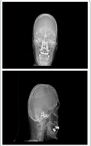
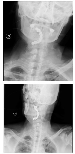
A sixty years old patient was admitted to our intensive rehabilitation department after a right nucleo-capsular hemorrhagic stroke. He was affected by hypertension, paroxysmal atrial fibrillation, recent infective pneumonia. The day after admission missing of upper fixed dental prosthesis was noticed during neurological evaluation. The patient was asymptomatic with normal oxygen saturation while breathing in ambient air. An urgent chest x-ray was performed. Displaced dental prosthesis at hypo-pharynx tract was noticed. Endoscopic extraction of the foreign body was performed without complications and the patient was able to continue his rehabilitative course.
Aspiration or ingestion of foreign bodies is not uncommon in the adult population [1]. It has been suggested that foreign body aspiration or swallowing is more frequent in selected groups including elderly, psychotics, alcohol/drug abusersand neurologic patients [2,3]. The more commonly swallowed foreign bodies among adults are fish bones (9–45%), bones other than fish bones (8–40%), and dentures (4–18%) [3]. The delay in diagnosis can result in significant morbidity. Ifundetected, retained foreign bodies can lead to significantedema that causes obstruction or perforation, or both [4]. Ingested foreign bodies progress through the digestive tract spontaneously in 80–90% of cases; however, 10–20% of patients require endoscopy for removal, and less than 1% undergo surgery [5].
References
- Haidary A, Leider JS, Silbergleit R. Unsuspected swallowing of a partial denture. Am J Neuroradiol. 2007; 28: 1734– 1735.
- Ramos MB, Fernández-Villar A, Rivo JE, et al. Extraction of airway foreign bodies in adults: Experience from 1987–2008. Interact Cardiovasc Thorac Surg. 2009; 9: 402– 405.
- Ambe P, Weber SA, Schauer M, Knoefel WT. Swallowed foreign bodies in adults. Dtsch Arztebl Int. 2012; 109: 869–875. https:// doi.org/10.3238/arztebl.2012.0869.
- Singh B, Kantu M, Har-El G, et al. Complications associated with 327 foreignbodies of the pharynx, larynx, and esophagus. Ann Otol Rhinol Laryngol. 1997; 106: 301– 304
- Magalhães Costa P, Carvalho L, Rodrigues JP, Túlio MA, Marques S, et al. Endoscopic management of foreign bodies in the upper gastrointestinal tract: An evidence-based review article. GE Port J Gastroenterol. 2015; 23: 142–152. https://doi.org/10.1016/j. jpge.2015.09.002.