
Journal of Clinical Images and Medical Case Reports
ISSN 2766-7820
Clinical Image - Open Access, Volume 3
Rare case of Marchiafava Bignami Disease (MBD)
Amarkak Abdillahi Wais1*; Daoud Mohamed Ali1; Madina Rabileh Yassin1; Wilson Bizimana1; Meriem Fikri1; Najwa Ech-Chrifel Kettani1; Mohamed Jiddane2; Firdaous Touarsa2
1 Department of Radiology, Hospital of Specialty, CHU Ibn-Sina, Mohamed V University, Rabat, Morocco.
2 Department of Radiology, Military Instructions Hospital Mohamed V. Mohamed V University, Rabat, Morocco.
*Corresponding Author: Amarkak Abdillahi Wais
Department of Radiology, Hospital of Specialty, CHU Ibn-Sina, Mohamed V University, Rabat Morocco.
Email: amarkak.a.wais@gmail.com
Received : Jan 31, 2022
Accepted : Mar 21, 2022
Published : Mar 28, 2022
Archived : www.jcimcr.org
Copyright : © Wais AA (2022).
Abstract
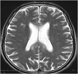
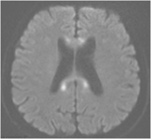
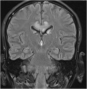
We report a case of a 45-year-old patient with a history of severe alcoholism who presented to the emergency department an impaired unconscious and weakness with upper and lower limbs. The patient was referred to our department for an MRI investigation. MRI showed abnormalities signal in the corpus callosum. The disease is due to a deficiency of each type of vitamin B, can be the result of necrosis and demyelination of the corpus callosum and later involves the genu and splenium.
Keywords: Marchiafava Bignami Disease; Corpus Callosum; MRI.
Citation: Wais AA, Ali DM, Yassin MR, Bizimana W, Fikri M, et al. Rare case of Marchiafava Bignami Disease (MBD). J Clin Images Med Case Rep. 2022; 3(3): 1760.
Description
Marchiafava-Bignami disease is a rare, toxic demyelinating disorder of the central nervous system associated with chronic alcoholism and malnutrition, essentially due to deficiency of vitamin B. The patient was presented an impaired consciousness and weakness with upper and lower limbs extremity. Radiology features a very important for the early diagnosis and management such as CT scan (Computed Tomography) and MRI (Magnetic Resonance Imaging). MRI images showed hyperintensity T2 weightedand T2 FLAIR with hyper intensity diffusion with low diffusion coefficient (ADC) in the corpus callosum, genu and splenium.
References
- Tian TY. Marchiafava Bignami disease. Stat Pearls. 2021.
- Carrilho PEM. Marchiafava Bignami disease: A rare entity with a poor outcome. Rev Bras Ter Intensiva. 2013; 25: 68–72.
- Ironside R. Central demyelination of the corpus callosum (MARCHIAFAVA-BIGNAMI DISEASE): With report of a second case in GREAT BRITAIN. Brain. 1961; 84: 212-230.