
Journal of Clinical Images and Medical Case Reports
ISSN 2766-7820
Case Report - Open Access, Volume 3
Pleural plaques
Moh’d Al-Halawani1; Yazan Abdeen2; Luise Froessl3*
1 Division of Pulmonary and Critical Care Medicine, SUNY Downstate Medical Center, Brooklyn NY 11203, USA.
2 Pulmonary Department, Saint Michael’s Medical Center, Seton Hall University School of Health and Medical Sciences, Newark, NJ 07102, USA.
3 Baylor College of Medicine, Houston, TX, USA.
*Corresponding Author: Luise Froessl
Medicine/Pulmonary, Baylor College of Medicine,
Houston, USA.
Email: luisefroessl@gmail.com
Received : Feb 25, 2021
Accepted : Mar 24, 2022
Published : Mar 31, 2022
Archived : www.jcimcr.org
Copyright : © Froessl L (2022).
Citation: Halawani MA, Abdeen Y, Froessl L. Pleural plaques. J Clin Images Med Case Rep. 2022; 3(3): 1769.
Description
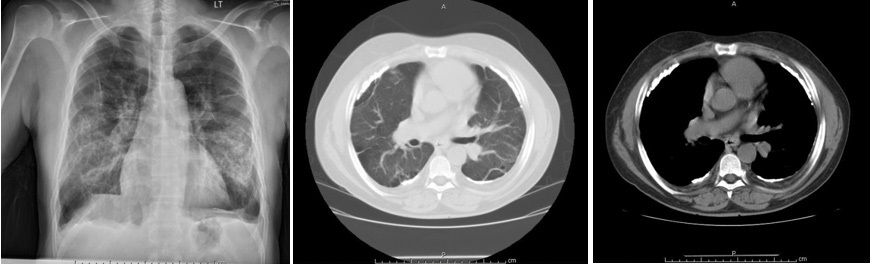
A 55-year-old male, with past history of COPD, presented with progressive shortness of breath of five days duration, associated with cough and increased sputum production. He had a 30 pack year smoking history and worked as a clerk in a plumbing store for two decades. On examination, he was afebrile, breathing at 20 /min with Oxygen saturation of 91% on 2 liters of oxygen. He was also tachycardic at 104 /min. Chest exam revealed fair air entry bilaterally with diffuse wheezing and a prolonged expiratory phase. Initial laboratory workup was within normal limits. His chest X-ray showed bilateral extensive opacity in the mid-lower zones, and hyperinflation. An axial CT scan was done (Figure 1).
What is the diagnosis?
The CT scan of the chest showed bullous emphysema and bilateral calcified pleural plaques. Pleural plaques are the most common pathologic pulmonary response to asbestos inhalation and often hallmark of the disease [1]. It affects approximately 50% of persons with heavy and prolonged exposure to asbestos and serves as evidence of asbestos inhalation. Plaques are usually symmetrical along the lateral chest wall and often not visible in Chest X-Ray, requiring CT scan [1]. This makes the above images unique in nature. Pleural Plaques are usually asymptomatic, but may lead to mild restrictive lung disease changes compared to those without pleural plaques yet exposed to asbestos. [2]. It is a benign finding that requires no treatment but several studies deem it as a risk factor for potential malignancy, making candidates with asbestos related pleural plaques, a high risk category for screening [3].
References
- Elshazley M, Shibata E, Hisanaga N, Ichihara G, Ewis AA, Kamijima M, et al. Pleural plaque profiles on the chest radiographs and CT scans of asbestos-exposed Japanese construction workers. Ind Health. 2011; 49: 626-633. Epub 2011.
- Kopylev L, Christensen KY, Brown JS, Cooper GS. A systematic review of the association between pleural plaques and changes in lung function. Occup Environ Med. 2014; oemed-2014-102468.
- Pairon JC, Andujar P, Rinaldo M, Ameille J, Brochard P, et al. . Asbestos exposure, pleural plaques, and the risk of death from lung cancer. Am J Respir Crit Care Med. 2014; 190: 1413-1420.