
Journal of Clinical Images and Medical Case Reports
ISSN 2766-7820
Clinical Image - Open Access, Volume 3
Roth’s spots in leukemia
Chia Chun Huang1; Hung Jen Chien2; Sung Yuan Hu1,3,4,5*
1 Department of Emergency Medicine, Taichung Veterans General Hospital, Taichung, Taiwan.
2 Department of Ophthalmology, Taichung Veterans General Hospital, Taichung, Taiwan.
3 School of Medicine, Chung Shan Medical University, Taichung, Taiwan.
4 Institute of Medicine, Chung Shan Medical University, Taichung, Taiwan.
5 Department of Post Baccalaureate Medicine, College of Medicine, National Chung Hsing University, Taichung, Taiwan.
*Corresponding Author: Sung-Yuan Hu
Department of Emergency Medicine, Taichung Veterans General Hospital, Taichung City, Taiwan.
Email: song9168@pie.com.tw
Received : Mar 14, 2022
Accepted : Apr 05, 2022
Published : Apr 12, 2022
Archived : www.jcimcr.org
Copyright : © Sung-Yuan H (2022).
Abstract
Roth’s spots are retinal hemorrhages with white or pale centers, which can be seen in numerous conditions. Ocular disorders have been described in 39.3% of patients with diagnosis of leukemia. Incidences of intraocular manifestations are 7.7% in patients of chronic leukemia. Mechanism of retinal hemorrhages is related to hyperviscosity syndrome caused by leukocytosisor direct infiltration. Fundoscopy is a mandatory examination by ophthalmologist in patients of leukemia with intraocular disorders. We present the case of Roth’s spots in a 32-year-old man of chronic myeloid leukemia.
Keywords: Leukemia; Retinal hemorrhages; Roth’s spots.
Abbreviations: CML: Chronic Myeloid Leukemia.
Citation: Huang CC, Chien HJ, Hu SJ. Roth’s spots in leukemia. J Clin Images Med Case Rep. 2022; 3(4): 1788.
Description
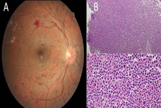
A 32-year-old man, with occasional dizziness and fatigue for one year, presented acute onset of central blurred vision of the left eye occurred. No focal neurologic signs neither limitation in eye of movement were found. Complete blood cells were white blood cells 417,700/mm3 , hemoglobin 8.6 g/dl, and platelet 273,000/mm3 . Lactate dehydrogenase 955 U/l and alkaline phosphatase 233 U/l and others were unremarkable. Fundoscopic examination revealed multiple Roth’s spots at fovea of the left eye (Figure 1A). Bone marrow biopsy demonstrated hyperplasia in myeloid series, blasts, increased megakaryocytes with dwarf size, and hypolobulated nucleus (Figure 1B), which was compatible with Chronic Myeloid Leukemia (CML). Resolution of CML was completed and visual acuity returned to normal status after chemotherapy for CML.
Roth’s spots are retinal hemorrhages with white or pale centers, which can be seen in numerous conditions, such as bacterial endocarditis, complications of leukemia, intracranial hemorrhage, vasculitis, opportunistic infections, hypertension, diabetes mellitus, sepsis, and effect of medical therapy [1-5]. Ophthalmic manifestations have been described in 39.3% of patients with diagnosis of leukemia [1,2]. Incidence of intraocular manifestations is 7.7% in CML [1]. Visual impairment is only present in 4% of patients at initial diagnosis of CML [2]. Roth’s spots account for a quarter of intraocular manifestations [1,3]. Roth’s spot is more common in acute (20.5%) than in chronic (4.9%) leukemia and its mechanism can be explained due to hyperviscosity syndrome caused by leukocytosis or direct infiltration [1-3,5]. Several studies reveal ocular disorders as the early sign of worsening clinical course with significantly poor prognosis in patients with CML [2]. Overall, it’s important that prompt detailed review of systems and laboratory evaluation when Roth’s spots have been noted. Fundoscopy is a mandatory examination by ophthalmologist in patients of leukemia with intraocular disorders [5].
Declarations
Conflict of interest declaration: None.
Consent: This study was approved by the Institutional Review Board of Taichung Veterans General Hospital (No. CE21215A).
Funding: This work was supported by grants from the Taichung Veterans General Hospital (Grant numbers: TCVGH1107202C). The funder had no role in the study design, data collection and analysis, decision to publish, or preparation of the manuscript.
References
- Dhasmana R, Prakash A, Gupta N, Verma SK. Ocular manifestations in leukemia and myeloproliferative disorders and their association with hematological parameters. Ann Afr Med. 2016; 15: 97-103.
- Ilo OT, Adenekan AO, Alabi AS, Onakoya AO, Aribaba OT, Kehinde MO, et al. Ocular manifestations of leukaemia: A teaching hospital experience. Niger Postgrad Med J. 2019; 26: 205-210.
- Buzaid AN, Al-Amri AM. Sudden visual Loss as an initial manifestation of chronic myeloid leukemia. Saudi J Med Med Sci. 2017; 5: 278-280.
- Varga Z, Pavlu J. Images in clinical medicine. Roth’s spots. N Engl J Med. 2005; 353: 1041.
- Němčanská S, Stepanov A, Němčanský J. Ophthalmic manifestations of acute leukaemias. Cesk Slov Oftalmol. 2018; 74: 98-101.