
Journal of Clinical Images and Medical Case Reports
ISSN 2766-7820
Short Report - Open Access, Volume 3
Imaging in vascular medicine: Right gastroepiploic artery aneurysm
Tobias Ribeiro dos Santos Neto1*; Jorge Luís Bezerra Holanda2; Carla Franco Costa Lima2; Mara Mônica Rocha Rolim2
1 Radiology Departament, Instituto Doutor José Frota, R. Barão do Rio Branco, 1816 - Centro, Fortaleza - CE, Brazil, 60025-061.
2 Radiology Departament, Hospital Geral de Fortaleza, R. Ávila Goularte, 900 - Papicu, Fortaleza - CE, Brazil, 60150-160.
*Corresponding Author: Tobias Ribeiro dos
Santos Neto
Integrated Laboratory of Biochemistry, Hatisaburo
Masuda, Center for Ecology and Socio-Environmental Development of Macaé (NUPEM), Campus UFRJ
Macaé, National Institute of Science and Technology in Molecular Entomology - INCT-EM, Macaé,
Brazil.
Email: tobiasribeiro26@gmail.com
Received : Mar 22, 2022
Accepted : Apr 21, 2022
Published : Apr 28, 2022
Archived : www.jcimcr.org
Copyright : © Santos Neto TR (2022).
Abstract
A 79-year-old woman was transferred to our service with a complaint of hematemesis associated with hypotension. There was no history of trauma. She was hemodynamically stable, but developed melena and anemia during hospitalization. She underwent abdominal ultrasound which showed an expansive oval formation in the gastroduodenal transition, determining compression of the intestinal lumen and liquid dilatation of the gastric chamber. There was intense flow in the Doppler study. Computed tomography with contrast confirmed the presence of a saccular aneurysmal dilatation of the right gastroepiploic artery with a mural thrombus, in close relationship with the stomach wall. This paper proposes to present a case report involving aneurysms of the splanchnic circulation, more specifically of the right gastroeploic artery, a relatively rare location for this pathology.
Citation: Santos Neto TR, Bezerra Holanda JL, Costa Lima CF, Rocha Rolim MM. Imaging in vascular medicine: Right gastroepiploic artery aneurysm. J Clin Images Med Case Rep. 2022; 3(4): 1811.
Case report
A 79-year-old woman was referred to our service with a complaint of hematemesis associated with hypotension. During the past year, there were reports of intermittent episodes of fever, diarrhea, abdominal pain, gastric fullness, and unmeasured weight loss. At the time of admission, she was hemodynamically stable, having evolved with melena and anemia.
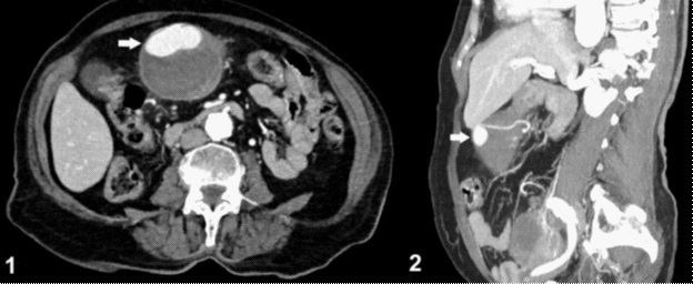
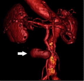
An abdominal Doppler ultrasound was performed, which showed an expansive oval formation in the gastroduodenal transition, determining compression of the intestinal lumen and liquid dilatation of the gastric chamber. The formation showed intense flow on Doppler study and was surrounded by hypoechoic material. In correlation, contrast-enhanced computed tomography of the abdomen confirmed the presence of a saccular aneurysmal dilatation of the right gastroepiploic artery with a mural thrombus, in close relationship with the stomach wall, measuring approximately 7.3 X 7.1 X 7.1 cm (Figure 1), even better demonstrated with the Volume Rendering Technique (VRT) reconstruction.
Discussion
Arterial aneurysms of the splanchnic circulation are relatively rare entities of multifactorial etiology, with atherosclerosis being the most prevalent cause. Congenital vascular abnormalities, infection, trauma, inflammation, secondary vasculitis in autoimmune connective tissue diseases and Segmental Arterial Mediolysis (SAM) are other possible causes.
The aneurysms were described in the following decreasing order of prevalence: splenic artery aneurysms (60%), hepatic artery (20%), superior mesenteric artery (5.5%), celiac artery (4.4%) and finally gastroepiploic artery (3%). Therefore, the right gastroepiploic artery aneurysm is among one of the rarest forms of presentation and, to date, there are no embracing reviews in the literature about this topic. This work aims to present a case report about this unusual pathology.
There is still no consensus on the pathophysiology of the disease, however the most plausible mechanism currently accepted is that there is a weakening of the vascular media, associated with an increase in intravenous pressure, which would be the predisposing factors for rupture.
Symptoms are usually nonspecific, such as vague abdominal pain. Because of this, most cases are diagnosed late either in an emergency laparotomy after aneurysm rupture or even during the autopsy. A clinical picture of severe, abrupt or rapidly progressive upper abdominal pain associated with signs of hemodynamic instability should lead to the suspicion of pain of probable vascular origin, especially if the patient has one or more of the aforementioned risk factors.
Imaging exams are essential in the diagnosis, surgical planning and monitoring of aneurysms. The exams of choice are abdominal ultrasound, computed tomography (CT) and angiography. In the emergency context, tomography with intravenous contrast is the exam of preference, because with it, in addition, is possible to detect the focus of active bleeding, in some cases.
On ultrasound, aneurysms usually appear as circumscribed anechoic tubular enlargements and can be fusiform or saccular. By Doppler analysis, the turbulent internal flow can be visualized. At CT, the attenuation coefficient of the lumen of the aneurysm follows that of another arterial lumen in the non-contrast images and in all contrasted phases, if it is patent. Pseudoaneurysms may have an image aspect similar to that of aneurysms, however their margins tend to be more irregular and is surrounded by a hematoma.
Recognition and treatment should be performed quickly regardless of the size of the aneurysm, given the high probability of rupture, which can approach 90% in cases of gastroepiploic artery aneurysms. In cases where there is hemodynamic instability, supportive therapy with volume replacement is used (saline solution and blood products, when necessary). Surgical excision can be laparoscopic or laparotomy, the latter being the treatment of choice in emergency cases. The endovascular approach with embolization by transarterial catheter aims at hemostasis and may be an option right after angiography in those individuals with hemorrhage with risk of death.
References
- Talha Sarigoz, Sedat Carkit, Omer Topuz, Tamer Ertan, Ali Koc. Spontaneous rupture of right gastroepiploic artery aneurysm: a rare cause of hemorrhagic shock. Case report. Sao Paulo Med. J. vol.136 no.5 São Paulo Sept./Oct. 2018 Epub Aug 21, 2017.
- Ahmad S. Ashrafi, Michael J. Horkoff, Junaid Yusuf, Alfred Stedman. Ruptured gastroepiploic artery aneurysm: A case report. Int J Surg Case Rep. 2017; 41: 132-133.
- Yuki Murakami, Hiroaki Saito, Shota Shimizu, Yusuke Kono, Hirohiko Kuroda, Tomoyuki Matsunaga, et al. A Case of Unruptured Right Gastroepiploic Artery Aneurysm Successfully Resected by Laparoscopic Surgery. Yonago Acta Med. 2017; 60(1): 56-58.
- S.J.Goa, S.G.Leea, M.S.Leea, B.S.Choa, M.K.Leea, Y.J.Choia, C.N.Kima, Y.J.Kanga, J.S.Parka, H.Y.Hanb. Ruptured Gastroepiploic Artery Aneurysm as an Unusual Cause of Hemoperitoneum. European Journal of Vascular and Endovascular Surgery. 2014; 48(3): 355.
- Robert A. Jesinger, MD, MSE, Andrew A. Thoreson, MD, Ramit Lamba, MBBS, MD. Abdominal and Pelvic Aneurysms and Pseudoaneurysms: Imaging Review with Clinical, Radiologic, and Treatment Correlation. RadioGraphics 2013; 33: E71-E96. Published online 10.1148/rg.333115036.