
Journal of Clinical Images and Medical Case Reports
ISSN 2766-7820
Clinical Image - Open Access, Volume 3
Osteogenesis imperfecta XV: An ultra-rare genetic disease with neurological anomalies
Erlane Marques Ribeiro1,2*; Raffaela Montálverne2; Andre Luiz Santos Pessoa1,3; Pablo Coimbra4
1 Hospital Infantil Albert Sabin, Brazil.
2 Faculdade de Medicina Unichristus, Brazil.
3 Faculdade de medicina da Universidade Estadual do Ceará, Brazil.
4 Hospital Geral de Fortaleza, Brazil.
*Corresponding Author: Erlane Marques Ribeiro
Rua Cesar Fonseca 455/202 B; CEP 60.192260,
Bairro Cocó; Fortaleza-Ceará-Brazil.
Email: erlaneribeiro@yahoo.com.br
Received : Mar 30, 2022
Accepted : Apr 25, 2022
Published : May 02, 2022
Archived : www.jcimcr.org
Copyright : © Ribeiro EM (2022).
Keywords: Osteogenesis imperfecta; Brain stem; Hydrocephalus; WNT1 protein; Human.
Citation: Erlane MR, Raffaela M, Andre LSP, Pablo C. Osteogenesis imperfecta XV: An ultra-rare genetic disease with neurological anomalies. J Clin Images Med Case Rep. 2022; 3(5): 1815.
Clinical image description
A six-year-old Brazilian boy was born to non-consanguineous and healthy parents at 40 weeks of gestation via cesarean delivery. The family history is unremarkable. He had diagnosis of hydrocephalus since prenatal period and the treatment was the placement of a shunt at the first week of life. Because he had many bony fractures in 50 days of life, he became bisphosphonate therapy. The phenotype of osteogenesis imperfecta has been characterized by multiple fractures but the neurological abnormalities are unusual [1]. Intravenous bisphosphonate therapy is still the most widely used drug treatment approach [1,2].
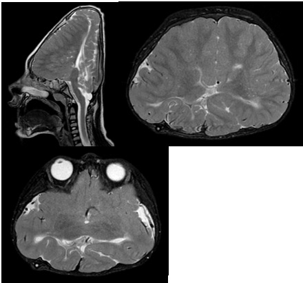
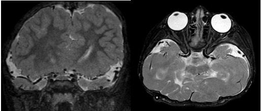
We found significant neurological malformations similar it was seen in the literature [3]. Brain MRI revealed alterations in the supratentorial parenchyma with a diffuse alteration of the sulcation pattern, corpus callosum of difficult characterization, focal communication between the white matter of the cerebral hemispheres through the midline, reduction of the dimensions of the lateral ventricles, alteration of the signal in T2/FLAR in the supratentorial white matter in the periventricular regions with reduction of the parenchymal mantle, volumetric reduction of the brain stem, notably of the pons and signs that appear fusion of the thalamus with important reduction of the dimensions of the cerebellum. There is only a small remnant of the superior aspect of the cerebellar parenchyma. Only one artery was characterized in the topography of the anterior cerebral arteries, malformation of the basal ganglia, diffuse thinning of the optic nerves, and there was also a reduction in the thickness of the optic chiasm (Figures 1 and 2).
the supratentorial parenchyma with a diffuse alteration of the sulcation pattern, corpus callosum of difficult characterization, focal communication between the white matter of the cerebral hemispheres through the midline, reduction of the dimensions of the lateral ventricles, alteration of the signal in T2/FLAR in the supratentorial white matter in the periventricular regions with reduction of the parenchymal mantle, volumetric reduction of the brain stem, notably of the pons and signs that appear fusion of the thalamus with important reduction of the dimensions of the cerebellum. There is only a small remnant of the superior aspect of the cerebellar parenchyma. Only one artery was characterized in the topography of the anterior cerebral arteries, malformation of the basal ganglia, diffuse thinning of the optic nerves, and there was also a reduction in the thickness of the optic chiasm (Figures 1 and 2).
References
- Panigrahi I. Novel mutation in a family with WNT1-related osteoporosis. European Journal of Medical Genetics [Internet]. 2018; 61(7): 369-371. Available from: https://www.sciencedirect. com/science/article/abs/pii/S1769721217303385?via%3Dihub DOI: 10.1016/j.ejmg.2018.01.017.
- Tauer JT, Robinson M-E, Rauch F. Osteogenesis Imperfecta: New Perspectives from Clinical and Translational Research. JBMR1 Plus. 2019; 3(8): 1-10.
- Aldinger KA, et al. Variable brain phenotype primarily affects the brainstem and cerebellum in patients with osteogenesis imperfecta caused by recessive WNT1 mutations. J Med Genet [Internet]. 2016. [cited 2020; 53(6): 427-430. Available from: https://www.ncbi.nlm.nih.gov/pmc/articles/PMC4898782/. DOI: 10.1136.