
Journal of Clinical Images and Medical Case Reports
ISSN 2766-7820
Case Report - Open Access, Volume 3
Successful anaesthetic management of case of tessier cleft with left encephalocele: A Case Report
Debendra Kumar Tripathy 1 ; Vaishali Gupta 2 ; Ruhi Sharma 3 *; Abhishek BV 2
1 Department of Anesthesiology, Additional Professor, AIIMS, Rishikesh, Uttrakhand, India.
2 Department of Anesthesiology, Junior Resident, AIIMS, Rishikesh, Uttrakhand, India.
3 Department of Anesthesiology, Senior Resident, AIIMS, Rishikesh, Uttrakhand, India.
*Corresponding Author: Ruhi Sharma
Department of Anesthesiology, Senior Resident,
AIIMS, Rishikesh, Uttrakhand, India.
Email: drsharma.ruhi@gmail.com
Received : Apr 04, 2022
Accepted : Apr 29, 2022
Published : May 06, 2022
Archived : www.jcimcr.org
Copyright : © Ruhi Sharma (2022).
Citation: Debendra KT, Vaishali G, Ruhi S, Abhishek BV. Successful anaesthetic management of case of tessier cleft with left encephalocele: A case report. J Clin Images Med Case Rep. 2022; 3(5): 1823.
Introduction
Craniofacial clefts, estimated to be between 1.4 and 4.9 per 100,000 live births [1], are extremely rare congenital malformations. Apart from functional, psychosocial, and aesthetic effects on a patient’s life, these anomalies pose a great challenge to anesthesiologists in managing the airway of patients. We here describe the case report of successful management of Tessier cleft with left encephalocele.
Case report
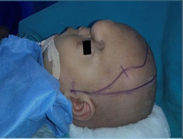
A 7 month old infant, weighing 8 kg, was diagnosed with Tessier cleft with left meningoencephalocele and proposed for cranioplasty with left box osteotomy. The anomaly was associated with hypertelorism. Birth history and antenatal course were found to be insignificant. Developmental milestones were reported appropriate for age. Systemic examination and routine blood examination were within normal limits. No associated cleft lip or palatal defects were seen. There were no other associated genetic abnormalities. NCCT head showed left frontal calvarial defect with herniation of CSF and dura mater showing meningoencephalocele. Anaesthetic preparation was done according to standard guidelines. After taking written informed consent from the parents and confirming nil per oral status, the patient was taken inside the operation theatre and all ASA standard monitors were attached. Preoxygenation was done and anesthesia was induced with injection fentanyl 15mcg+ injection propofol 25 mg. After check ventilation and confirming ability to ventilate, injection atracurium 4mg was given for muscle relaxation followed by intubation with size 4.0 RAE south pole endotracheal tube. Bilateral air entry was checked and confirmed with ETCO2 monitoring and the tube was fixed at the 12 cm mark. Packing of the throat was performed. The patient was mechanically ventilated to maintain normocarbia. In addition to standard ASA monitoring, left femoral artery was cannulated for invasive blood pressure monitoring. Central venous line was also secured in the left femoral vein. Maintenance of anesthesia was done with oxygen, air, sevoflurane (MAC 0.8- 1.2), and intermittent boluses of injection atracurium (0.1 mg/ kg). Other drugs given were injection fentanyl 15 mcg, injection paracetamol 120 mg, injection dexamethasone 1.5 mg, injection hydrocortisone 16 mg. Blood products and crystalloid transfusion was done in calculated doses keeping an eye over blood loss and urine output. Intraoperative arterial blood gas analysis and blood glucose were within normal limits. Total duration of surgery was 8 hours with an intraoperative blood loss was 150 ml. The intraoperative course was uneventful and the child was shifted to PICU with ET in-situ for further management. The patient was extubated on POD1. Postoperative blood investigations and arterial blood gas analysis were normal. The intensive care unit course was uneventful and the child was shifted to ward on POD3 and was discharged on POD 10.
Discussion
The palatal shelves and eventually the hard and soft palate are formed through 50th -60th gestational days. Failure of proper fusion of these structures leads to formation of facial clefts [2]. These account for the second most common congenital malformation of the entire body. Most of the classifications of craniofacial clefts are restricted to analysis of sharply defined areas of face. Morian [3] gave the first classification of craniofacial anomalies in 1886. Subsequently Sanvenero-Rosselli [4], Burian [5], and other authors attempted classification of these defects. Tessier [6] in 1990 gave a simplified and widely accepted classification. He classified craniofacial defects according to the anatomical defects as facial, craniofacial, and laterofacial [7]. The etiology of the craniofacial cleft is multifactorial. Maternal infections such as toxoplasmosis, and intake of thalidomide drugs, alteration in phenylalanine levels have been implicated in its causative factors [8]. Aetiopathogenesis includes genetic and environmental factors that are correlated with changes in transmembrane protein1, gad1, glutamate decarboxylase. Advanced maternal age, stress, malnutrition, smoking, alcohol, and drug abuse [2] have been associated with increased incidence. Airway problems are the major anesthetic concerns in craniofacial anomalies. Therefore, to recognize the potential difficulties, careful preoperative examination and selection of the appropriate technique needs to be done [9]. The potential anesthetic complications, in this case, were anticipated difficult bag and mask ventilation, difficult intubation due to associated encephalocele. Anticipating difficult airway, the difficult airway cart had been prepared with different types and sizes of face masks, oral airways, adequate sizedsupraglottic airways including igel and classic LMA. Tracheostomy set and fibreoptic bronchoscope were kept ready to aid intubation. Inability to ventilate constitutes the most common cause of airway-related morbidity in the pediatric population [2]. We were able to achieve adequate seal and proper mask ventilation using size 1 Rendell-baker-soucek mask attached to Jackson rees circuit. We used a neuromuscular blocking agent only afterconfirming the check ventilation and laryngoscopy. Face and scalp are highly vascular areas of the body, so surgeries of these areas are associated with high risk of bleeding. Volume loss due to blood loss and long duration of surgery are potential causative factors for hypothermia, metabolic acidosis and associated coagulopathy. Proper planning and anticipation of such events were kept in mind while preparing for surgery. Near ambient temperature was maintained in the operating room and warm fluids were used throughout the surgery to prevent hypothermia. Intraoperative larger fluid shifts were anticipated so blood and blood products were kept ready. Close monitoring for ETCO2, urine output, and all hemodynamic parameters were done to recognize any changes at the earliest. Intravascular volume was maintained with crystalloids and blood products keeping an eye on blood loss, third space loss, urine output, and other insensible losses. The causes for deterioration can be airway obstruction, hypoxia, hypercarbia, metabolic acidosis, coagulopathy, hypothermia, cerebral edema, intracranial bleeding, hypoglycemia, and electrolyte imbalance [2]. The postoperative ICU ventilatory bed was kept ready in view of anticipated problems. Surgery was uneventful and followed by an uneventful postoperative course and the child was discharged successfully.
Conclusion
Although airway problems are a major challenge in craniofacial anomalies but careful examination, anticipating difficulties and proper preparation and a team approach can lead to successful management.
References
- Kalantar-Hormozi A, Abbaszadeh-Kasbi A, Goravanchi F, Davai NR. Prevalence of Rare Craniofacial Clefts. J Craniofac Surg. 2017; 28(5): e467-70.
- Baruah U, Dayal M, Giridhar K, Virender. Successful management of a case of Tessier’s cleft number 0 and 14 with associated encephalocoele. Indian J Anaesth. 2016; 60(8): 597-9.
- Kawamoto HK. The kaleidoscopic world of rare craniofacial clefts: order out of chaos (Tessier classification). Clin Plast Surg. 1976; 3(4): 529-72.
- Sanvenero-Rosselli G. Developmental pathology of the face and the dysraphic syndrome; - an essay of interpretation based on experimentally produced congenital defects. Plast Reconstr Surg 1946. 1953; 11(1): 36-8.
- Burian F. Median clefts of the nose. Acta Chir Plast. 1960; 2: 180- 9.
- David DJ, Moore MH, Cooter RD. Tessier clefts revisited with a third dimension. Cleft Palate J. 1989;26(3):163-84; 184-185.
- Tessier P. Anatomical classification facial, cranio-facial and latero-facial clefts. J Maxillofac Surg. 1976; 4(2): 69-92.
- Kumar K, Ninan S, Saravanan P, Prakash KS, Jeslin L. Airway management in an infant with tessier N. 4 anomaly. J Anaesthesiol Clin Pharmacol. 2011; 27(2): 239-40.
- Carenzi B, Corso RM, Stellino V, Carlino GD, Tonini C, Rossini L, et al. Airway management in an infant with congenital centrofacial dysgenesia. Br J Anaesth. 2002; 88(5): 726-8.