
Journal of Clinical Images and Medical Case Reports
ISSN 2766-7820
Clinical Image - Open Access, Volume 3
A leaking food pipe: Bronchoesophageal fistula secondary to lung cancer
Ibiyemi Oke1*; Arpan Pokhrel1; Shefali Amin1; Kim James2
1Department of Internal Medicine, Reading Hospital, Tower Health System, West Reading, Pennsylvania, USA.
2Pulmonology and Critical Care Department, Reading Hospital, Tower Health System, West Reading, Pennsylvania, USA.
*Corresponding Author : Ibiyemi Oke
Department of Internal Medicine, Tower Health
System, Reading Hospital, 301 S 7th Avenue, West
Reading, PA, 19611, USA.
Email: Ibiyemi.oke@towerhealth.org
Received : May 16, 2022
Accepted : Jun 08, 2022
Published : Jun 15, 2022
Archived : www.jcimcr.org
Copyright : © Oke I (2022).
Citation: Oke I, Pokhrel A, Amin S, James K. A leaking food pipe: Bronchoesophageal fistula secondary to lung cancer. J Clin Images Med Case Rep. 2022; 3(6): 1890.
Case presentation
A 58-year-old female with advanced lung cancer, hypertension, hyperlipidemia, pulmonary artery thrombosis, bilateral subclavian artery stenosis, and vocal cord dysfunction presented to the ER with cough, shortness of breath, dysphagia, and weight loss. HR 114 bpm, RR 24/min, SpO2 95% on 4L, Temperature 36.2o C. She was lethargic, chronically ill-looking, and had bronchial breath sounds and crackles in the right upper lobe. Chest x-ray revealed enlarging previously known right upper lobe mass and loculated right pleural effusion. Contrast CT of the chest showed unchanged necrotic right apical mass, increased consolidation inferior to a mass in the right middle lobe, and metastatic infiltration of the right lateral 6th rib. She also had purulent secretions in all airways on bronchoscopy. Shortly after bronchoscopy, she had an episode of aspiration and required Intensive Care Unit (ICU) admission.
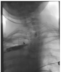
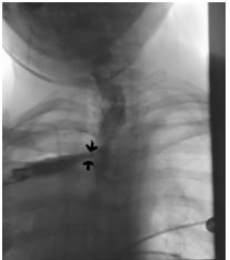
On her second day in the ICU, a repeat chest x-ray showed complete opacification of the right hemithorax, suspected to be due to mucus plugging. There was an improvement with mucolytics and chest physiotherapy. During a Videofluoroscopic Study (VFS), she was coughing and became more hypoxemic. Barium was noted in the tracheobronchial tree, with drainage through a large fistula between the esophagus and the right upper lobe cavity.
The patient required mechanical ventilation, pressors, and intravenous antibiotics. She was not a good surgical candidate due to her poor clinical status. Acquired Bronchoesophageal Fistula (BEF) is a potentially life threatening complication of esophageal and bronchial malignancy [1]. It may result from the primary malignancy or its management. BEF increases the risk of recurrent lung infection, sepsis, and death. Barium esophagography is the gold standard for diagnosing BEF, but it may also be seen on radiographs and during Endoscopy [2]. Patients that qualify for intervention may benefit from esophageal stents, prostheses, or bypass surgery. Our patient’s clinical status precluded any intervention.
References
- Kleinberg LR, Brock MV, Jagannath SB, et al. Abelof MD, Armitage JO, Niederhuber JE, et al. Cancer of the esophagus. Abeloff’s Clinical Oncology. Philadelphia, PA Churchill Livingstone, An Imprint of Elsevier. 2008; 1399-1429.
- Zhang BS, Zhou NK, Yu CH. Congenital bronchoesophageal fistula in adults. World J Gastroenterol. 2011; 17: 1358-1361.