Journal of Clinical Images and Medical Case Reports
ISSN 2766-7820
Clinical Image - Open Access, Volume 3
Choroidal melanoma in the setting of nevus of ota
Muhamad Festok1,2; Behnam Rabiee1,2*; Michael Gaspari1,2; Imtiaz Chaudhry1,2
1Trinity Health Mid-Atlantic, Nazareth Hospital, Department of Ophthalmology, Philadelphia, PA, USA.
2IC Laser Eye Care, Bensalem, PA, USA.
*Corresponding Author : Behnam Rabiee
Research Faculty, Department of Ophthalmology,
Trinity Health Mid-Atlantic, Nazareth Hospital,
Philadelphia, PA, USA.
Phone: +1 (215) 639-4500;
Email: Behnam.Rabiee@gmail.com
Received : May 20, 2022
Accepted : Jun 20, 2022
Published : Jun 27, 2022
Archived : www.jcimcr.org
Copyright : © Behnam Rabiee (2022).
Citation: Festok M, Rabiee B, Gaspari M, Chaudhry I, et al. Choroidal melanoma in the setting of nevus of ota. J Clin Images Med Case Rep. 2022; 3(6): 1911.
Clinical image description
The patient is a 56 years old female who presented to the office with a complaint of blurry vision OS for a few weeks. Upon initial encounter, several pigmented macules were visible around the left eye and on the lids. The patient mentioned she had these lesions since birth.
The vision was 20/20 OD and 20/25 OS. No relative afferent pupillary defect was present. External ocular movements were within normal limits. The slit-lamp exam showed pigmentedmacules on the eyelid skin, palpebral and bulbar conjunctivae, consistent with a Nevus of Ota.
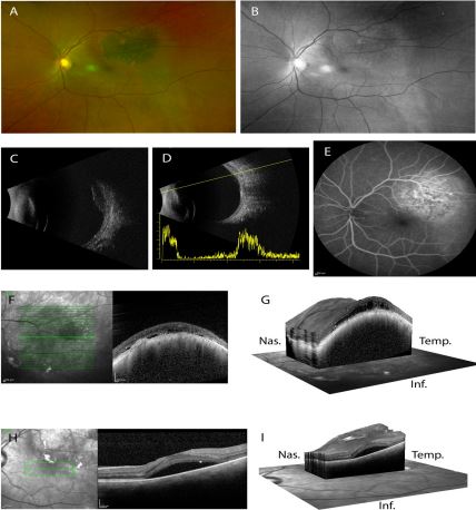
B-Scan (Figure 1C) showed a raised homogenous lesion with an apical diameter of 3.28 mm, and choroidal shadowing.
A-Scan (Figure 1D) showed low to medium reflectivity of the lesion.
Fundus fluorescein angiography showed initial blocking and late staining and pooling in different areas of the lesion, as well as a double circulation pattern (Figure 1E).
Fundus OCT with 2D and 3D reconstruction (Figure 1F, G) showed a heterogenous dome-shaped lesion with subretinal fluid, shaggy photoreceptors, and thinning of the overlying choriocapillaris as well as optical shadowing.
Macular OCT with 2D and 3D reconstruction (Figure 1H, I) showed subretinal fluid in the macula, adjacent to the domeshaped lesion.
These findings in a patient with a history of Nevus of Ota are consistent with a diagnosis of Choroidal Melanoma. Nevus of Ota is a well-known risk factor for this disease, and patients with a Nevus of Ota should have regular retinal examinations to catch the possible melanoma early and proceed with life-saving treatment.