
Journal of Clinical Images and Medical Case Reports
ISSN 2766-7820
Case Report - Open Access, Volume 3
Forgotten double J stent with maximum stone burden
Shah Aamir1*; Khan Abrar2; Raina Aamir1; Wani Inzamam2
1Department of Radiology, Sher-e-kashmir Institute of Medical sciences, (SKIMS) SOURA, J&K, INDIA.
2Department of Surgery, Sher-e-kashmir Institute of Medical sciences, (SKIMS) SOURA, J&K, INDIA.
*Corresponding Author : Shah Aamir
Department of Radiology, Sher-e-kashmir Institute
of Medical sciences, (SKIMS) SOURA, J&K, INDIA.
Email: : shaaamer@gmail.com
Received : May 23, 2022
Accepted : Jun 22, 2022
Published : Jun 29, 2022
Archived : www.jcimcr.org
Copyright : © Aamir S (2022).
Abstract
The Double J stent, or DJ stent, is a self-retaining ureteral stent that is widely used in urologic practice. The primary goal of the DJ stent is to enable adequate kidney drainage into the urinary bladder. However, because of widespread use, lack of patient education, or due to a lack of adherence to regular follow-up, patients may end up with a forgotten DJ stent, which can stay undiagnosed in the pelvi-ureteral system for years and cause a lot of complications before coming to attention. In addition to increasing patient morbidity, mortality, and healthcare costs, it can also pose legal issues for the practitioner. We present a unique case of repetitively neglected (forgotten) DJ Stent in a 28-year old male who had the stent placed 11 years back as a part of Percutaneous Nephrolithotomy (PCNL) for right renal stone, had forgotten it for 6 years, underwent open cystolithotomy for the encrusted DJ stent and concomitant urinary bladder stone, was again lost to follow up, and presented back 5 years later with an encrusted DJ stent with large bladder calculus and calcular deposits along the entire length of the stent. The patient was then managed in two sittings, as an open cystolithotomy, followed a few months later by a combination of uretroscopic lithotripsy and percutaneous lithotomy. To our knowledge, this study reports the forgotten stent with the maximum stone burden available in the literature. This case presents a unique challenge to the operating surgeon, as the entire entity had to be removed in two different sittings and involved a combination of open and endoscopic procedures.
Keywords: Forgotten DJ stent; Encrustations; Management.
Citation: Aamir S, Abrar K, Aamir R, Inzamam W, et al. Forgotten double J stent with maximum stone burden. J Clin Images Med Case Rep. 2022; 3(6): 1915.
Background
The Double J Stent, a self-retaining ureteral stent used for renal drainage, is widely used in urologic practice. It is used as a part of routine uretroscopy for stone disease, after a multitude of reconstructive surgeries to allow for uretral healing, for obstructive uropathy, before Extracorporeal Shock Wave Lithotripsy (ESWL), for obstructive anuria, etc [1]. However, serious complications such as encrustation, migration, stone formation, fragmentation, and infection can be seen if the stents have been placed for a long time [2]. Damiano et al. observed flank pain in 25.3%, irritative bladder symptoms in 18.8%, hematuria in 18.1%, and fever in 12.3% of the patients [3]. Patients who are asymptomatic are more likely to ignore or forget about their stent [4]. The incidence of encrustation over the DJ stent increases as the length of time the stent is in place increases [5]. The literature suggests that the DJ stent generally needs to be replaced or removed within 6 weeks to 6 months [6].
Case presentation
A 28-year-old male presented to us with a complaint of intermittent pain in the right flank region since 5 months. On eliciting a detailed history from the patient, the past medical history revealed that he had undergone right-sided Percutaneous Nephrolithotomy (PCNL) with the placement of a Double J Stent for right renal stone about 11 mm in size in the right renal pelvis 10 years ago (year 2011) in another hospital. However, the patient was lost to follow-up for stent removal and had presented to the previous hospital’s OPD as a case of forgotten right-sided Double J Stent after 5 years (Year 2016) with the plain X-ray kidney, ureter, and bladder (X-ray KUB) suggestive of encrustation of Double J Stent involving almost the whole length of the right ureter and right pelvicalyceal system. The patient underwent open cystolithotomy in 2016 for the same at the previous hospital. The bladder stone was removed, but due to the fixity of the Double J stent at the upper end, the stent could not be brought down and was cut at the lower end. The patient was again lost to follow up and has now presented to our OPD in 2020. On examination, the patient was afebrile. Blood reports showed no evidence of leukocytosis. Urea and serum creatinine were 43 and 1.19, respectively. All other blood reports and urinalysis were normal.
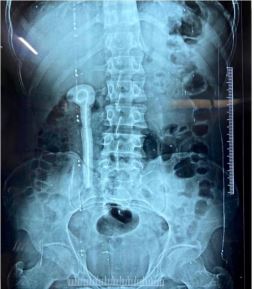
A plain X-ray of the kidney, ureter, and bladder (X-ray KUB) was done, which revealed a large urinary baldder stone measuring approximately 7.3 cm in the largest diameter with encrusted Double J stent and calculus deposits along the entire length of the stent measuring approximately 25 cm in length (Figure 1).
Abdominal ultrasonography was done, which showed a right-sided Double J Stent in situ with localised hydronephrosis and medullary renal disease in the right kidney and grade I splenomegaly.
It was followed by a DTPA (Diethylenetriamine Pentaacetate) scan reporting a poorly visualised right kidney with hydronephrosis and grossly decreased function, a GFR of 23.3 and a differential function of 27.4, whereas the left kidney had a dilated pelvic-calyceal system and extrarenal pelvis with adequate washout, a GFR of 61.3 and a differential function of 72.6.

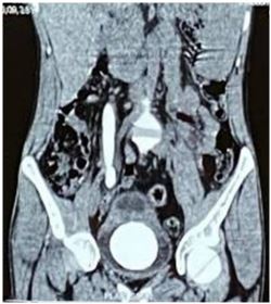
Non-Contrast-Computed Tomography of the Kidney Ureter Bladder (NCCT-KUB) showed evidence of G III hydronephrosis with mild stranding around the renal pelvis in the right kidney and the upper end of the Double J Stent in situ. There was a dilated right ureter with a Double J stent in situ with evidence of calculus deposits around the stent extending up to the renal pelvis. The urinary bladder had a 7.3 x 6.1 cm calculus (620 HU) and the lower end of a Double J Stent in situ. The left kidney and ureter were normal (Figure 2).
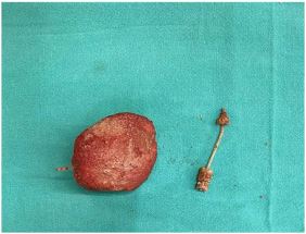
Subsequently, a treatment decision was taken on the basis of the clinical presentation and radiographs of the patient. He was operated on as a case of open cystoloithotomy in our hospital on March 17, 2021. A pfannensteil incision was made and around 8 cm of calculus was retrieved from the bladder. An attempt to retrieve the whole Double J Stent was made, but only the lower half could be retrieved. The upper half, due to its fixity, could not be removed (Figure 3)
Post-operative X ray KUB showed completely cleared urinary bladder calculus as well as the lower half of the Double J stent

and its encrustations. There was residual encrusted Double J stent and accompanying calculus deposits in the renal pelvis and upper half of the ureter (Figure 4).
The patient was then discharged in a stable condition with upper half of Double J Stent with accompanying stone in situ, with a plan to remove it in the next sitting.
The patient was then admitted again for the residual encrusted Double J Stent and surrounding stone deposits. On June 2, 2021, right-sided Percutaneous Nephrolithotomy (PCNL) with Right-Sided Uretroscopic Lithotripsy (URSL) was done in our hospital. Using a rigid uretroscope, the residual right-sided DJ stent with heavy stone material encrustation along its entire length was visualized. It was fragmented, and the fragments were evacuated. It was followed by introducing a nephroscope with a 24 Fr dilatation in the right posterior midcalyx and the stone was visualized. It was also fragmented, and fragments were removed. Plain X-ray of the kidney, ureter, and bladder (Xray KUB) showed clear residual encrusted Double J stent and accompanying calculus deposits in the renal pelvis and upper half of the ureter (Figure 5). The patient developed hypotension in the postoperative period, which was managed by vasopressors and antibiotics. He was discharged in a stable condition.
Discussion
If forgotten, DJ stents, which are indispensible in urological procedures, pose a major challenge to the attending surgeon and to the patient as well. A stent with an indwelling time period of more than 3–6 months can be termed “forgotten” if not intended by the treating doctor [7]. The reasons behind a forgotten or retained stent can be attributed to inadequate counselling by the treating doctor and poor compliance on the part of the patient and his or her family [8]. Various factors that have been linked with the formation of encrustations and stone on a stent, such as long indwelling time, urinary sepsis, history of stone disease, chemotherapy, pregnancy, chronic renal failure, and metabolic and congenital abnormalities [9]. There have been extensive studies on the composition and the risk factors of encrustations. Calcium oxalate (43.8%), especially in its monohydrate form, constitutes most of the encrustation [10]. The incidence of encrustations is less with silicone DJ stents compared to polyurethane stents [10,11].
The clinical presentation of a forgotten DJ stent includes flank pain, irritative voiding symptoms, hematuria, pyrexia, stent fragmentation, and migration. In a study by Hao et al [12]. The most prevalent symptom was hematuria, which was followed by discomfort and bladder irritation.
The management and intervention depend on the preoperative status of the patient, location and size of stone and encrustations as well as severity of encrustations. Stent migration and fragmentation are important factors in determining the course of intervention. For the management of mild encrustation, several studies have reported the role of ESWL followed by retrograde extraction of the DJ stent. In patients with moderate-tosevere encrustations and stone presence, modalities such as transurethral Cystolithotomy (CLT), ureteroscopy, and PCNL are used [13,14]. It has been suggested in a number of studies that the distal part of the stent should be removed first, followed by the proximal end [15]. Patient counselling regarding the DJ stent by the treating doctor is very important. Patient compliance is also important and is reflected by the quality of counselling provided by the treating urologist. Maintaining a simple stent registry can achieve almost 98% of DJ stent removal at the due date, which reduces morbidity associated with encrusted stent removal and anaesthetic drugs. Sabharwal et al. reported a computer-based stent registry with a patient-directed automated information system, and it sends automated SMS initially, followed by letters in case they fail to respond; however, a long-term prospective trial is needed for its efficacy [16].
Conclusion
A forgotten DJ stent with encrustations and stone burden is a serious urological problem for the patient and treating doctor. They are a major source of major burden on patients in terms of increasing morbidity and additional procedures that can be simply avoided if the stents are removed in time. Hence, it is of utmost importance that the patients and their attendants be counselled and informed about the presence of a stent in the patient’s system after such procedures, and also be explained in detail about the hazards of a forgotten stent. Good practise should include mentioning the presence of DJ Stent in bold letters on their discharge certificates and also keeping a log of patients’ names, residences, and contact numbers in a separate hospital or departmental database, so as to ensure timely follow-up and removal.
Declarations
Consent for publication: Informed Consent taken from Patient for Publication of Clinical Images.
Competing interests: None.
Funding: None.
References
- Aboutaleb HA, Ali TA, Gawish M, Omar MK. Fluoroscopy-free double-J stent placement through ureteroscope working channel postuncomplicated ureteroscopic laser lithotripsy: A novel technique Urol Ann. 2019; 11: 39-45.
- anohar CS. 2016; 30: A106. http://ovidsp.ovid.com/ovidweb.cgi?T=JS&PAGE=reference&D=emex&NEWS=N&AN= 613823925 (Suspected Torsion Score in Patients Presenting with Acute scrotum.J Endourol).
- R Damiano, A Oliva, C Esposito, M De Sio, R Autorino, M D’Armiento, et al. Early and late complications of double pigtail ureteral stent Urol Int. 2002; 69: 136-140.
- Abdelaziz AY, Fouda WB, Mosharafa AA, Abelrasoul MA, Fayyad A, Fawzi K, et al. Forgotten ureteral stents: Risk factors, complications and management. African J Urol. 2018; 24
- El-Faqih SR, Shamsuddin AB, Chakrabarti A, Atassi R, Kardar AH, Osman MK, et al. Polyurethane internal ureteral stents in treatment of stone patients: Morbidity related to indwelling times. J Urol. 1991; 146: 1487–1491.
- Borboroglu PG, Kane CJ. Current management of severely encrusted ureteral stents with a large associated stone burden. J Urol. 2000; 164: 648–650.
- Agarwal S, Sarpal R, Pathak P, Biswas M, Mittal A, Rathore K, et al. Tricks and tacks in the management of the forgotten double J stent. Int Surg J. 2018; 26: 792.
- Monga M, Klein E, Castañeda-Zúñiga WR, Thomas R, et al. The forgotten indwelling ureteral stent: A urological dilemma. J Urol. 1995; 153: 1817.
- Sohrab A, Aneesh S, Sureka SK, Varun M, Nitesh P, Manoj K, et al. Forgotten reminders: An experience with managing 28 forgotten double-j stents and management of related complications. Indian J Surg. 2015; 77: 1165–1171.
- Bouzidi H, Traxer O, Doré B, Amiel J, Hadjadj H, Conort P, et al. Characteristics of encrustation of ureteric stents in patients with urinary stones.
- Mardis HK, Kroeger RM, Morton JJ, Donovan JM. Comparative evaluation of materials used for internal ureteral stents. J Endourol. 1993; 7: 105–115.
- Hao P, Li W, Song C, Yan J, Song B, Li L, et al. Clinical evaluation of double-pigtail stent in patients with upper urinary tract diseases: Report of 2685 cases. J Endourol. 2008; 22: 65–70.
- Milicevic S, Bijelic R, Jakovljevic B. Encrustation of the ureteral double J stent in patients with a solitary functional kidney – A case report. Med Arch. 2015; 69: 265–268.
- Singh I, Gupta NP, Hemal AK, Aron M, Seth A, Dogra PN, et al. Severely encrusted polyurethane ureteral stents: Management and analysis of potential risk factors. Urology. 2001; 58: 526– 531.
- Weedin JW, Coburn M, Link RE. The impact of proximal stone burden on the management of encrusted and retained ureteral stents. J Urol. 2011; 185: 542–547.
- Sabharwal S, Macaden AR, Abrol N, Mukha RP, Kekre NS, et al. A novel computer based stent registry to prevent retained stents: Will patient directed automated short message service and letter generator help? Indian J Urol. 2014; 30: 150–152