
Journal of Clinical Images and Medical Case Reports
ISSN 2766-7820
Clinical Image - Open Access, Volume 3
Clinical images: Cinematic rendering of tophaceous gout
Hannah S Recht1*; Elliot K Fishman2
1Northwestern University Feinberg School of Medicine, Department of Radiology, 676 North St. Clair Street, Suite 800. Chicago, IL 60611, USA.
2Johns Hopkins University School of Medicine, The Russell H Morgan Department of Radiology and Radiological Science, 601 North Caroline Street, JHOC 4260 C, Baltimore, Maryland 21287, USA.
*Corresponding Author : Hannah S Recht, MD,
Northwestern University Feinberg School of Medicine, Department of Radiology, 676 North St. Clair
Street, Suite 800, Chicago, IL 60611, USA.
Tel: (216) – 570-2406;
Email: Hannah.recht@northwestern.edu
ORCID ID: 0000-0003-2697-3831
Received : May 24, 2022
Accepted : Jun 24, 2022
Published : Jul 01, 2022
Archived : www.jcimcr.org
Copyright : © Recht HS (2022).
Keywords: Three-dimensional imaging; Gout; Musculoskeletal radiology
Citation: Recht HS, Fishman EK. Clinical images: Cinematic rendering of tophaceous gout. J Clin Images Med Case Rep. 2022; 3(7): 1922.
Descriptions
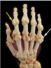
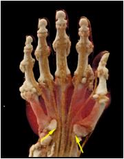
A 77 year-old-man with a complex medical history, notable for multiple myeloma in remission and gout, presented for noncontrast Computed Tomography (CT) of the right upper extremity using dual energy technique, from which cinematic rendered images were created (Figure 1). The cinematic rendered images provide a unique, photorealistic, three-dimensional representation of the extensive tophi and their relationship to the adjacent structures within the hand.
Gout is a common inflammatory arthropathy caused by the deposition of monosodium urate crystals. Multiple advanced imaging techniques have been utilized in the evaluation of gout, including dual-energy CT [1,2]. However cinematic rendering, through its use of a complex global lighting model, has the unique ability to provide three-dimensional photorealistic images from volumetric CT datasets. Cinematic rendering can help improve visualization of complex anatomy and pathology, particularly for those without formal training. The photorealistic images created by this novel technique may help increase patient understanding and improve communication between patients and clinicians.
Declarations
Funding information: Hannah S Recht: None.
Elliot K Fishman: Receives grant funding from GE Healthcare and Siemens, and is a founder and stockholder, HipGraphics.
Declarations of interest: None
References
- Desai MA, Peterson JJ, Garner HW, Kransdorf MJ. Clinical utility of dual-energy CT for evaluation of tophaceous gout. Radiographics. 2011; 31: 1365-1375; discussion 1376-1377.
- Girish G, Glazebrook KN, Jacobson JA. Advanced imaging in gout. AJR Am J Roentgenol. 2013; 201: 515-525.