
Journal of Clinical Images and Medical Case Reports
ISSN 2766-7820
Research Article - Open Access, Volume 3
Evaluation of phytochemical constituents and antimicrobial activities of ethyl acetate and butanol fractions of Periploca aphylla Decne
Umbreen Rashid1,2,3*; Muhammad Rashid Khan1; Jasia Bokhari1; Shumaila Jan1
1Department of Biochemistry, Faculty of Biological Sciences, Quaid-i-Azam University, Islamabad, Pakistan.
2Department of Microbiology, Faculty of Biological Sciences, Quaid-i-Azam University, Islamabad, Pakistan.
3Department of Life Sciences, Abasyn University, Islamabad, Pakistan.
*Corresponding Author : Umbreen Rashid
Department of Microbiology, Quaid-i-Azam University, Islamabad, Pakistan.
Email: umbreen.rashid@gmail.com
Received : Jun 14, 2022
Accepted : Jul 12, 2022
Published : Jul 19, 2022
Archived : www.jcimcr.org
Copyright : © Rashid U (2022).
Abstract
Introduction: Plants have been utilized for the cure of different diseases since ancient times. Medicinal importance of the plants is due to presence of phytochemicals having specific physiological action on the human body. A wide variety of activities have been found in these phytochemicals which might help in preventing chronic disorders. Periploca aphylla Decne belongs to the family Asclepiadoideae, is traditionally used for the cure of cerebral fever and as stomachic.
Objectives: In this study, phytochemical constituents and antimicrobial potentials of ethyl acetate and butanol fractions of crude methanolic extract of P. aphylla were investigated.
Methodology: Qualitative determination of Phytochemical constituents (tannins, saponins, flavonoids, cardiac glycosides, terpenoids, coumarins, phlobatanins and anthraquinones) in the extracts was performed using standard procedures. Antibacterial and antifungal activities were determined using agar well diffusion method and tube dilution method respectively.
Results: The phytochemical analysis of ethyl acetate and butanol fractions of P. aphylla crude methanol extract confirmed the existence of tannins, saponins, alkaloids, flavonoids, cardiac glycosides and terpenoids. Ethyl acetate fraction did not show any activity against the tested bacterial strains except K. pneumonia. Whereas butanol fraction was active against all the strains. Ethyl acetate fraction showed inhibition against A. fumigates and F. solani while did not show any inhibition against M. pyriformis. The n-butanol fraction showed inhibition against all the tested fungal strains.
Conclusion: It is evident from the above results that P. aphylla is a rich reservoir of phytochemical compounds which contributes to its various ethnomedicinal uses.
Keywords: Periploca aphylla; Phytochemicals; Ethyl acetate; Butanol; Antimicrobial.
Citation: Rashid U, Khan MR, Bokhari J, Jan S. Evaluation of phytochemical constituents and antimicrobial activities of ethyl acetate and butanol fractions of Periploca aphylla Decne. J Clin Images Med Case Rep. 2022; 3(7): 1956.
Introduction
Medicinal plants have been the source of medicine to all civilizations since long. Plants are selected for pharmacological analysis using one of the frequent methods i.e. ethnobotany [1]. Traditional medicine is employed as a major health care system in many developing countries [2,3]. The interest in maintaining personal health and safety by using medicinal plants is increasing due to bioprospecting of novel plant-derived drugs along with rising costs of prescribed medicines [4]. The large variety of bioactive molecules produced by plants, are mostly evolved as chemical defense against infection or predation, make them a rich source of medicine. About 74% of 119 pharmaceutical or biotechnology drugs derived from plants are used in modern medicine corresponding to their traditional uses [5,6]. Hence, ethnomedical data provides a better chance to find active plants rather than random approach [7,8].
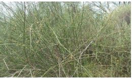
Periploca aphylla Decne. is a medicinally important plant. It belongs to the family Asclepiadoideae having 348 genera, with about 2,900 species. They are primarily located in the tropics to subtropics, particularly in Africa and South America. It is commonly found in the northern parts of Pakistan and has a variety of medicinal uses in the indigenous system of medicine [9].
It is locally named as “Bata”. The milky juice of P. aphylla is externally applied to swellings and tumors. It is also used for the treatment of cerebral fever and as stomachic [10]. Latex from its stem is used as chewing gum. The flowers are used as vegetables. Sometimes, they have been used for emetic purposes, expectorant, laxative, diuretic and for wart removal. There are a number of known triterpenes and steroids which have been isolated from this species [11].
There are several chemical substances which produce a definite physiological effect on human body and hence are responsible for the medicinal importance of plants. The most significant of these plant bioactive constituents includes phenolic acids, flavonoids, terpenoids, lignins, tannins, stilbenes, coumarins, alkaloids, quinones, betalains, amines, vitamins and other metabolites having antioxidant potential [12,13].
There has been a universal trend in recent times towards using natural phytochemicals found in teas, berry crops, herbs, vegetables, fruits and beans [14-16]. Moreover, natural antioxidant ingestion reduces the risks of cardiovascular disorders, cancer, diabetes and age related diseases [17,18]. The nature of drugs can be predicted by the preliminary phytochemical screening. It also helps to detect different constituents present in different polarity solvent.
The evaluation of medicinal plants on a large scale for different biological activities is the first essential step to isolate and characterize the active principles which will lead to drug development. Antimicrobial research is required for the discovery and growth of new therapeutic antimicrobial agents. This effort is encouraged due to lack of good antifungal agents and the ongoing trouble of development of resistance to existing antibacterial agents towards novelty [19]. In the past years, numerous reports are presented on the antibacterial activity of plant extracts against human pathogenic bacteria which showed that plants were significant source of potentially valuable structures to develop novel chemotherapeutic agents [20-27].
One of the important alternative approaches to manage diseases and control antibiotic resistance is the isolation of antibacterial active principles from higher plants. The drugs derived from plants are believed to cause less or no side effects in comparison to synthetic antibiotics [28]. In vitro studies have revealed a great deal of phytochemicals which belongs to different chemical classes to have inhibitory effects on all types of microorganisms [29].
Materials and methods
Plant material
Plant samples of Periploca aphylla were collected in April-May 2010 from Margalla Hills Islamabad, Pakistan. Further identified by Dr. Saleem Ahmad and deposited the voucher specimen at Herbarium of Pakistan Museum of Natural History, Islamabad (Voucher No. 069721). Whole plants were collected and shade dried at temperatures range of 21-30°C. Resulting dried samples were powdered using a blender and kept at room temperature in polythene bags. The extraction was carried out according to the following procedure.
Preparation of extract
Methanol extract: Dried powdered samples of P. aphylla (1.5 kg) were soaked in 3 litres of methanol (95%) for one week (3 times) at room temperature with random shaking and stirring. The filtration of extracts was carried out by using Whatman filter paper No. 42 (125 mm). After combining the resulting filtrates, they were concentrated at 40oC in a rotary vacuum evaporator to get a solid, gummy mass (PAME). The extract was stored at -4oC in airtight vials till further use.
Preparation of fractions: The methanol extract was partitioned using a liquid–liquid extraction technique and the resultant partitions i.e. ethyl acetate and n-butanol fractions were concentrated by means of a vacuum evaporator and were stored at -4oC in airtight vials till further analysis.
Determination of phytochemical constituents
The extracts were evaluated for the occurrence of flavonoids, tannins, saponins, phlobatannins, cardiac glycosides, alkaloids, terpenoids and anthraquinone using simple qualitative methods of Harborne [30], Trease and Evans [31] and Sofowora [32].
Salkowski test for terpenoids: To a 5 ml of extract/fraction; 2 ml of CHCl3 and 3 ml of H2SO4 (concentrated) were added. Terpenoid constituents were confirmed by the formation of a reddish brown colored interface.
Test for alkaloids: An amount of 0.1 g of extract and each fraction was warmed with 4 ml of 1% HCl followed by filtration. Titration of the filtrates (2 ml) was done individually with (a) Dragendroff’s reagent and (b) Mayer’s reagent. The presence of alkaloids was indicated by yellow or cream precipitate formation.
Test for saponin: About 0.1 g of the fraction was mixed with distilled water (10 ml) and then filtered after boiling in a water bath. An aliquot of 2.5 ml of distilled water was added to 5 ml of the filtrate and shake it vigorously. Emulsion was formed after mixing the froth with olive oil (3 drops) and shaken strenuously.
Test for phlobatannins: Boiling of 80 mg of crude extract and each fraction was carried out in 1% aqueous hydrochloric acid in a small tube. Formation of red precipitates showed the presence of phlobatannins.
Keller- Kiliani test (for deoxy sugars in cardiac glycosides): From the crude extract and each fraction, 5 ml of aqueous extract (10 mg/ml) was taken. Then 2 ml glacial acetic acid, 1 drop of FeCl3 solution and 1 ml concentrated H2SO4 was added to it. Brown ring was formed at interphase indicating the existence of cardiac glycosides.
Test for coumarins: About 0.1 g of extract and each fraction was taken in a test tube. Moistening of Filter paper was done with 1 N NaOH solution and it was used to cover the mouth of the tube. Test tube was placed in boiling water for few minutes. Filter paper was removed to examine under UV light. Yellow fluorescence indicates the presence of coumarins.
Test for anthraquinones: Briefly, 0.1 g of extract and each fraction was boiled in 5 ml of 1% HCl followed by filtration. To the filtrate 5 ml of benzene was added and 10 % NH4OH was added in the benzene layer after shaking. The colour in the alkaline phase was observed. Violet or red colour formation indicated the existence of anthraquinones.
Test for tannins: About 0.1 g of the extract and each fraction was mixed with five ml of H2O and filtered. Formation of blue black or brownish green color after treating filtrate with a 0.1% ferric chloride (few drops) indicated the presence of tannins.
Test for flavonoids: A portion of the aqueous filtrate of extract and each fraction was mixed with 5 ml of dilute NH3 solution and add concentrated H2SO4. A yellow coloration is observed if flavonoid compounds are present.
Biological assays
Anti-bacterial assay
Preparation of bacterial inoculums: Four isolated colonies were inoculated in the 30 ml nutrient broth and incubated for 24 h at 37oC so that the growth in the broth was equivalent with McFarland standard (0.5%).
Antibacterial activity: Antibacterial potential of crude extract derived fractions was investigated by agar well diffusion method [33] using nutrient agar medium. Exactly two grams of nutrient agar was dissolved in 100 ml of distilled water (pH 7.0) and was autoclaved. It was cooled down to 45oC. Then 100 ml of this media was seeded with 1 ml of inoculum having size of 106 CFU/ml as per McFarland standard and after proper homogenization 75 ml was poured into the petri plate of 14 cm diameter. For agar well diffusion method, eleven wells per plate were made by using a sterile cup-borer (8 mm). Extract and fractions at concentration of 25, 15, 12.5, 10, 7.5, 5, 3, 2 and 1 mg/ml were prepared in Dimethyl Sulfoxide (DMSO). The test compounds (100 μl) were poured into the wells and petri plates were then incubated at 37oC for 24 hours. Simultaneously, roxythromycin and cefixime were used as positive controls each having the concentration of 0.5 mg/ml. DMSO was used as a negative control as well as the dilution medium for the positive controls. The lowest concentration inhibiting growth was taken as the Minimum Inhibitory Concentration (MIC).
Antifungal activity
Agar tube dilution method was used to evaluate antifungal activity [34]. The stock solution of extract and positive control; terbinafine were prepared at a concentration of 12 mg/ml by dissolving them in DMSO. As a negative control DMSO was used. 6.5 g of sabouraud dextrose agar was dissolved in 100 ml of distilled water (pH 5.6) to make media for fungus. Then 4 ml of it was added into screw cap tubes. These tubes were autoclaved for 15 min at 120°C and cooled down to 45°C. The stock solution (66.6 μl) was mixed with media to get the final concentration of 200 μg/ml of sabouraud dextrose agar. Then the tubes were solidified in the slanted position at 25°C. An agar surface streak was used by inoculating each tube with a piece of inoculum (4 mm diameter) taken from a seven days old culture of fungi. After seven days of incubation at 28 ± 1°C, visual observation of fungal growth inhibition was made. The % inhibition of growth was calculated with reference to the negative control.
Results
Qualitative studies of phytochemicals
Qualitative phytochemical screening was carried out on all the considered fractions of P. aphylla to identify the phytochemical classes, i.e. tannins, saponins, flavonoids, cardiac glycosides, terpenoids, coumarins, phlobatanins and anthraquinones.
Qualitative screening of P. aphylla: The phytochemical analysis of ethyl acetate and butanol fractions of P. aphylla crude methanol extract confirmed the existence of tannins, saponins, alkaloids, flavonoids, cardiac glycosides and terpenoids. However coumarins, phlobatanins and anthraquinones were absent in both fractions (Table 1).
Table 1: Phytochemical constituents of fractions of P. aphylla methanol extract.
| Phytochemicals | Extract/Fractions | |
|---|---|---|
| PAEE | PABE | |
| Tannins | + | + |
| Saponins | + | + |
| Flavonoids | + | + |
| Alkaloids | + | + |
| Cardiac glycosides | + | + |
| Terpenoids | + | + |
| Phlobatannins | - | - |
| Coumarins | - | - |
| Anthraquinone | - | - |
PAEE: P. aphylla ethyl acetate fraction;
PABE: P. aphylla n-butanol fraction.
Anti-bacterial activity
The antibacterial potential of ethyl acetate and butanol extracts of P. aphylla were tested against five strains of bacterial test organisms. The antibacterial activity of the extracts and their potency were quantitatively assessed by determining Minimum Inhibitory Concentration (MIC) values. The extracts were tested at various concentrations (1-25 mg/ml), and the evaluated MIC values are reported in Table 2.
MIC value of P. aphylla: Ethyl acetate fraction did not show any inhibition against M. luteus. n-butanol fraction exhibited the activity against M. luteus with MIC values of 15 mg/ml. Both the fractions showed activity against K. pneumonia as follows: n-butanol (MIC=5 mg/ml) and ethyl acetate (MIC=12.5 mg/ml). Only the n-butanol fraction was active (MIC=3 mg/ml) against the B. bronchiseptica and E. aerogenes (MIC=7 mg/ml). Ethyl acetate fraction did not show any activity against P. aeruginosa as well while n-butanol fraction showed MIC value of 15 mg/ml (Table 2).
Anti-fungal activity
Ethyl acetate and butanol fractions of P. aphylla were screened in vitro for their antifungal activity against three strains. The growth inhibition was measured and presented in Table 3.
Anti-fungal activity of P. Aphylla: Inhibition in growth of A. fumigates was recorded for n-butanol (21.05%) and ethyl acetate (16.66%).
The n-butanol fraction showed 50% inhibition against F. solani, while the ethyl acetate showed 18.75% inhibition. Low activity was shown by n-butanol (6.25%) against M. pyriformis whereas ethyl acetate fraction was unable to produce any inhibition in growth (Table 3).
Table 2: The MIC values of fractions of P. aphylla methanol extract.
| Bacterial strains | Minimum inhibitory concentration (mg/ml) | |||
|---|---|---|---|---|
| PAEE | PABE | Rox | Cef | |
| M. luteus | - | 15 | 0.06 | 0.05 |
| K. pneumoniae | 12.5 | 5 | 0.17 | 0.13 |
| B. bronchiseptica | - | 3 | 0.14 | 0.16 |
| E. aerogenes | - | 7 | 0.16 | 0.15 |
| P. aeruginosa | - | 15 | 0.14 | 0.12 |
-: not active; PAEE: P. aphylla ethyl acetate fraction; PABE: P. aphylla n-butanol fraction; Rox: Roxithromycin; Cef: Cefixime.
Table 3: Antifungal activity of fractions of P. aphylla methanol extract.
| Fungal Strains | Percent inhibition (%) | |||
|---|---|---|---|---|
| Negative | Positive | PAEE | PABE | |
| Aspergillus fumigates | 0 | 100 | 16.66 | 21.05 |
| Fusarium solani | 0 | 100 | 18.75 | 50 |
| Mucor pyriformis | 0 | 100 | 0 | 6.25 |
PAEE: P. aphylla ethyl acetate fraction; PABE: P. aphylla n-butanol fraction
Discussion
Plants have been utilized for the cure of different diseases since ancient times. The medicinal uses of plants growing throughout the world lie in the active constituents having direct action in the body. They are used in conventional as well as herbal medicine. Hence mankind is provided with a valuable gift by nature in the form of herbal drugs [35]. Medicinal importance of the plants is due to presence of phytochemicals having specific physiological action on the human body. A wide variety of activities have been found in these phytochemicals which might help in preventing chronic disorders. Alkaloids have been used for medicinal purposes since long [36]. Besides cytotoxicity, they have been reported to have antispasmodic, analgesic [30,37] and antibacterial properties [38,39]. The phytochemical analysis of P. aphylla has also revealed to contain saponins. Saponins are major ingredients in Chinese traditional medicine [40]. They have been acknowledged to inhibit inflammation, have antibiotic potential and defend against hypercholesterolemia [41]. Saponins can coagulate and precipitate red blood cells and are involved in central nervous system activities. Moreover, they can form foams in aqueous solutions and possess hemolytic, bitterness and cholesterol binding properties [39,42]. Thus these characteristics of saponins and their presence in the plant extracts tend to validate the use of P. aphylla in traditional medicine. Several triterpenoids isolated from plants displayed good analgesic and anti-inflammatory properties [43,44]. Some of the pentacyclic triterpenoids are used in medicine because of their broad range of biological activities including cytotoxic, anti-bacterial, anti-fungal, anti-viral, anti-inflammatory, analgesic, anti-allergic and anti-cancer [45]. Tannins have been used to treat ulcerated or inflamed tissues and they have significant activity in the prevention of cancer [46,47]. Tannins interfere with protein synthesis and bind to proline rich protein. Thus phytotherapeutically, the plants containing tannins are used to treat throat and mouth inflammations, skin injuries and nonspecific diarrhea [48]. Since tannins can precipitate proteins, they protect the underlying layers by acting as waterproof to the external layers of the exposed tissues. Moreover, tannins are also known to possess different physiological effects such as antisecretolytic, anti-irritant, antiphlogistic, antiparasitic and antimicrobial activities [49]. One of the ubiquitous and leading groups of plant metabolites are phenolic compounds [50]. The biological properties of phenolic compounds include antiatherosclerosis, antiinflammation, endothelial function improvement, anticancer, antiapoptosis, cardiovascular protection, cell proliferation and angiogenesis inhibition activities [51]. The medicinal plants rich in phenolic compounds have been reported to have antioxidant properties in many studies [52]. The natural antioxidants derived from the plants are mostly in the form of phenolic compounds for instance phenolic acids, flavonoid, tocopherols [53]. Phenols have been useful in the preparation of cresol and dettol which are antimicrobial compounds. The antioxidative properties and use of P. aphylla in different diseases are due to the presence of these phenolic compounds. Flavonoids produced by plants in reaction to microbial infection are hydroxylated phenolic compounds and they have shown antimicrobial activity against broad range of microorganisms in in vitro studies. This activity is might be due to their ability to form complexes with bacterial cell wall and extracellular soluble proteins [54]. Flavonoids show anticancer activities and are effective antioxidants [55]. Flavonoids have been found to act by effecting membrane permeability and inhibiting membrane-bound enzymes like phospholipase A2 and ATPase [56], and the antioxidant action mechanism of P.aphylla may be explained by this property. Moreover, flavonoids as a result of their anion radicals serve as health promoting compounds [57]. The results found in our study imply that these phytochemical compounds might be the bioactive constituents in P. aphylla thus making these plants an important reservoir of medicinally important bioactive compounds. The plants provide potentially valuable structures in order to develop novel chemotherapeutic agents. In order to achieve this target, in vitro antibacterial assay is a first step [20]. A number of reports are accessible during the past years on the antibacterial potential of plant extracts against human pathogenic bacteria [21-27]. The synthetic antimicrobials are frequently coupled with side effects, whereas the plant derived antimicrobials having vast therapeutic potential can serve the rationale with less adverse effects [58]. Therefore, there is a requirement to constantly explore plant derived antimicrobials. More research is required to identify and resolve the full scale efficiency of antibacterial compounds from these plants. According to our results, the plant extracts were active against Gram negative as well as Gram positive bacteria. This might be an indication to the presence of wide range antibiotic compounds or general metabolic toxins, in addition to the pharmacological active metabolites such as flavonoid glycosides [59], spirostanol and furostanol saponins [60], amides and phytoterols [61]. Antimicrobial potential of the plants is perhaps due to high percentage of phenolics as both are always linked together [62]. It can also be due to the presence of different phytochemical constituents for example flavonoids, terpenoids, alkaloids, saponins and tannins in the plant extracts. The toxicity of saponins to parasite worms (anthelmintic activity), insects (insecticidal activity), fish (piscidal activity), molluscs (molluscicidal), their antibacterial, antifungal and antiviral activities are well known [63,64]. The membranolytic properties of saponins are involved in their antibacterial effects instead of simply changing the surface tension of extracellular medium, therefore get affected by population density of microbes [65]. Flavonoids, and tannins present in the extract have been associated with antimicrobial effects in various studies using plant extracts [66]. Plant derived alkaloids are usually found to have antimicrobial potential [67]. Alkaloids may be helpful against AIDS associated intestinal infections [68] as well as HIV infection [69]. Persistent opportunistic fungal infections have turned out to be a main factor for mortality and morbidity in immunocompromised patients [70]. The common fungal infections are mostly caused by the Candida and Aspergillus species. Recent trends in epidemiology have pointed out a shift towards infection caused by Aspergillus and non-albicans Candida [71].
Conclusion
The range of antimicrobial activities showed by these extracts could perhaps be explained by the presence of flavonoids, tannins, alkaloids and saponins. The microbes may inhibit more potently by purified components. Our results signify the potential of P. aphylla as medicinal agent’s sources, which may give leads in the constant exploration for antimicrobial agents from plants. Further studies on the purification of bioactive components and phytoconstituents can disclose the precise potential of the plant to restrain numerous pathogenic microorganisms. Thus, antimicrobial activity displayed by the extracts against different bacterial and fungal strains that are linked with different infectious diseases, may provide scientific validation for the ethnomedicinal uses of these plants.
Declarations
Conflict of interest: The authors declare no conflict of interest.
Funding: This research work is funded by Higher Education Commission of Pakistan (Indigenous Scholarship, Batch IV).
References
- Cox PA, Balick MJ. The ethnobotanical approach to drug discovery. Scientific American. 1994; 270: 60-65.
- Fransworth NR. Ethnopharmacology and future drug development. The North American experience. Journal of Ethnopharmacology. 1993; 38: 137-143.
- Houghton PJ. The role of plants in traditional medicine and current therapy. Journal of Alternative and Complementary Medicine. 1995; 1: 131-143.
- Sharma V, Sharma A, Kansal L. The effect of oral administration of Allium sativum extracts on lead nitrate induced toxicity in male mice. Food Chemistry and Toxicology. 2010; 48: 928-936.
- Barrett B, Kiefer D, Rabago D. Assessing the risks and benefits of herbal medicine: An overview of scientific evidence. Alternative Therapies in Health and Medicine. 1999; 5: 40-49.
- Newman DJ, Cragg GM, Snader KM. Natural products as sources of new drugs over the period 1981-2002. Journal of Natural Products. 2003; 66: 1022-1037.
- Lee KH. Novel Antitumor Agents from Higher Plants. Joun Wiley & Sons, Inc. Med Res Rev. 1999; 19: 569-596.
- Montbriand MJ. Herbs or natural products that decrease cancer growth. Oncology Nursing Forum. 2004; 31: 75-90.
- Shastri BN. Wealth of India. New Delhi, Sree Saraowaty. 1966; 313.
- Baquar SR. Medicinal and Poisonous plants of Pakistan. Karachi: Printas Press; 1989.
- Mustafa G, Anis E, Ahmed S, Anis I, Ahmed H, et al. Lupene-type triterpenes from Periploca a phylla. Journal of natural products. 2000; 63: 881-883.
- Zheng W, Wang SY. Antioxidant activity and phenolic compounds in selected herbs. Journal of Agricultural and Food Chemistry. 2001; 49: 5165-5170.
- Cai YZ, Sun M, Corke H. Antioxidant activity of betalains from plants of the Amaranthaceae. Journal of Agricultural and Food Chemistry. 2003; 51: 2288–2294.
- Kitts DD, Yuan YV, Wijewickreme AN, Hu C. Antioxidant properties of a North American gingseng extract. Molecular and Cellular Biochemistry. 2000; 203: 1-10.
- Wang SY, Jiao H. Correlation of antioxidant capacities to oxygen radical scavenging enzyme activities in blackberry. Journal of Agricultural and Food Chemistry. 2000; 48: 5672- 5676.
- Muselik J, Garcia Alonso M, Martin Lopez MP, Zelmieka M, Rivas Gonzalo JC, et al. Measurement of antioxidant activity of wine catechins, procyanidins, anthocyanins and piranoantocyanins. International Journal of Molecular Sciences. 2007; 8: 797-809.
- Ashokkumar D, Mazumder UK, Gupta M, Senthilkumar GP, Selvan VT, et al. Evaluation of antioxidant and free radical scavenging activities of Oxystelma esculentum in various in vitro models. Journal of Complementary and Integrative Medicine. 2008; 5.
- Veerapur VP, Prabhakar KR, Parihar VP, Kandadi MR, Ramakrishana S, et al. Ficus racemosa stem bark extract: A Potent antioxidant and a probable natural radioprotector. Evidence Based Complementary and Alternative Medicine. 2009; 6: 317-324.
- Silver L, Bostian K. Screening of natural products for antimicrobial agents. European Journal of Clinical Microbiology and Infectious Diseases. 1990; 7: 455-461.
- Tona L, Kambu K, Ngimbi N, Cimanga K, Vlietinck AJ, et al. Antiamoebic and phytochemical screening of some Congolese medicinal plants. Journal of Ethnopharmacology. 1998; 61: 57-65.
- Samy RP, Ignacimuthu S. Antibacterial activity of some folklore medicinal plants used by tribals in Western Ghats in India. Journal of Ethnopharmacology. 2000; 69: 63-71.
- Palombo EA, Semple SJ. Antibacterial activity of traditional medicinal plants. Journal of Ethnopharmacology. 2001; 77: 151-157.
- Kumaraswamy Y, Cox PJ, Jaspars M, Nahar L and Sarker SD, et al. Screening seeds of Scottish plants for antibacterial activity. Journal of Ethnopharmacology. 2002; 83: 73-77.
- Stepanovic S, Antic N, Dakic I, Svabicvlahovic M. In vitro antimicrobial activity of propilis and antimicrobial drugs. Microbiological Research. 2003; 158: 353-357.
- Bylka W, Szaufer Hajdrych M, Matalawska I, Goslinka O, et al. Antimicrobial activity of isocytisoside and extracts of Aquilegia vulgaris L. Letters in Applied Microbiology. 2004; 39: 93-97.
- Behera SK, Misra MK. Indigenous phytotherapy for genito-urinary diseases used by the Kandha tribe of Orissa, India. Journal of Ethnopharmacology. 2005; 102: 319-325.
- Govindarajan R, Vijayakumar M, Singh M, Rao CV, Shirwaikar A, et al. Antiulcer and antimicrobial activity of Anogeissus latifolia. Journal of Ethnopharmacology. 2006; 106: 57-61.
- Shariff N, Sudarshana MS, Umesha S, Hariprasad P. Antimicrobial activity of Rauvolfia tetraphylla and Physalis minima leaf and callus extracts. African Journal of Biotechnology. 2006; 5: 946-950.
- Cowan MM. Plant products as antimicrobial agents. Clinical Microbiology Reviews. 1999; 12: 564-582.
- Harborne JB. Phytochemicals Methods. London. Chapman and Hall Ltd. 1973; 49-188.
- Trease GE, Evans WC. Pharmacognosy. (11th Ed.) Bralliar Tridel Can. Macmillan Publishers. 1989.
- Sofowara AE. Medicinal plants and traditional medicine in Africa. 2nd ed. Ibadan, Nigeria. Spectrum books Ltd. 1993; 289.
- Bagamboula CF, Uyttendaele M, Debevere J. Antimicrobial effect of spices and herbs on Shigella sonnei and Shigella flexneri. Journal of Food Protection. 2003; 66: 668–673.
- Duraipandiyan V, Ignacimuthu S. Antibacterial and antifungal activity of Flindersine isolated from the traditional medicinal plant, Toddalia asiatica (L.) Lam. Journal of Ethnopharmacology. 2009; 123: 494–498.
- Saxena P, Arora A, Dey S, Malhotra Y, Nagarajan K, Singh PK, et al. Review on different methods to assess the antioxidant activity of some common plants of Indian traditional medicine. Journal of Drug Delivery and Therapeutics. 2011; 1: 36-39.
- Nobori T, Miurak K, Wu DJ, Takabayashik LA, Carson DA, et al. Deletion of cyclindependent kinase-4 inhibitor gene in multiple human cancers. Nature. 1994; 46: 753-756.
- Antherden LM. Textbook of Pharmaceutical Chemistry. 8th edition. London, UK. Oxford University Press. 1969; 813-814.
- Stray F. The Natural Guide to Medicinal herbs And Plants. London. Tiger Books International. 1998; 12-16.
- Okwu DE, Okwu ME. Chemical composition of Spondias mombin linn. plant parts. Journal of Sustainable Agriculture and the Environment. 2004; 6: 140-147.
- Liu J, Henkel T. Traditional Chineese Medicine (TCM): Are polyphenols and saponins the key ingredients triggering biological activities? Current Medicinal Chemistry. 2002; 9: 1483-1485.
- Just MJ, Recio MC, Giner RM, Cuellar MJ, Manez S, et al. AntiInflammatory activity of unusual lupine saponins from Bupleurum fruticescens. Planta Medica. 1998; 64: 404-407.
- Sodipo OA, Akiniyi JA, Ogunbamosu JU. Studies on certain characteristics of extracts of bark of Pansinystalia macruceras (K schemp) picrre Exbeille. Global Journal of Pure and Applied Sciences. 2000; 6: 83-87.
- Fernandez MA, De las Heras B, Garcia MD, Saenz MT, Villar A. New insights into the mechanism of action of the anti inflammatory Triterpene lupeole. Journal of Pharmacy and Pharmacology. 2001; 53: 1533-1539.
- Ismaili H, Sosa S, Brkic D, Fkih Tetouani S, IIidrrissi A, et al. Topical anti-inflammatory activity of extracts and compounds from Thymus broussonettii. Journal of Pharmacy and Pharmacology. 2002; 54: 1137-1140.
- Patocka J. Biologically active pentacyclic triterpenes and their current medicine signification. Journal of Applied Biomedicine. 2003; 1: 7-12.
- Motar MLR, Thomas G, Barbosa Fillo JM. Effects of Anacardium occidentale stem bark extract on in vivo inflammatory models. Journal of Ethnopharmacology. 1985; 95: 139-142.
- Ruch RJ, Cheng SJ, Klaunig JE. Prevention of cytotoxicity and inhibition of intercellular communication by antioxidant catechins isolated from Chinese Green tea. Carcinogens. 1989; 10: 1003-1008.
- Westendarp H. Effects of tannins in animal nutrition. Dtsch Tierarztl Wochenschr. 2006; 113: 264-268.
- Lutterodt GD, Ismail A, Basheer RH, Baharudin HM. Antimicrobial effects of Pisidium guajava extracts as one mechanism of its antidiarrhoeal action. Malaysian Journal of Medical Sciences. 2005; 6: 17-20.
- Singh R, Singh SK, Arora S. Evaluation of antioxidant potential of ethyl acetate extract/fractions of Acacia auriculiformis A. Cunn. Food and Chemical Toxicology. 2007; 45: 1216-1223.
- Han X, Shen T, Lou H. Dietry polyphenols and their biological significance. International Journal of Molecular Sciences. 2007; 8: 950-988.
- Mandal S, Patra A, Samanta A, Roy S, Mandal A, et al. Analysis of phytochemical profile of Terminalia arjuna bark extract with antioxidative and antimicrobial properties. Asian Pacific Journal of Tropical Biomedicine. 2013; 3: 960- 966.
- Ali SS, Kasoju N, Luthra A, Singh A, Sharanabasava H, et al. Indian medicinal herbs as source of antioxidants. Food Research International. 2008; 41: 1-15.
- Marjorie C. Plant products as antimicrobial agents. Clinical Microbiology Reviews. 1996; 12: 564-582.
- Akinmoladun AC, Ibukun EO, Afor E, Akinrinlola BL, Onibon TR, et al. Chemical constituents and antioxidant activity of Alstonia boonei. African Journal of Biotechnology. 2007; 6: 1197-1201.
- Li HB, Wong CC, Cheng KW, Chen F. Antioxidant properties in vitro and total phenolic contents in methanol extracts from medicinal plants. LWT-Food Science and Technology. 2008; 41: 385-390.
- Hausteen B. Flavonoids, a class of natural products of high pharmacological potency. Biochemical Pharmacology. 1983; 32: 1141-1148.
- Iwu M, Angela RD, Chin OO. New antimicrobials of plant origin. A reprint from Journal of Janicked perspective on New Crops and New Uses. Alexander: ASHS Press; 1999.
- Saleh NA, Ahmed AA, Abdalla MF. Flavonoid glycosides of Tribulus pentandrus and T. terrestris. Phytochemistry. 1982; 21: 1995-2000.
- Kostova I, Dinchev D. Saponins in Tribulus terrestris-chemistry and bioactivity. Phytochemistry Reviews. 2005; 4: 111-137.
- Xu YX, Chen HS, Liang HQ, Gu ZB, Lui WY, et al. Three new saponins from Tribulus terrestris. Planta Medica. 2000; 66: 545-550.
- Ravikumar S, Ramanathan G, Subhakaran M, Inbaneson SJ. Antimicrobial compounds from marine halophytes for silkworm disease treatment. International Journal of Medicine and Medical Sciences. 2009; 1: 184-191.
- Lacaille Dubois M, Wagner H. A review of the biological and pharmacological activities of saponins. Phytomedicine. 1996; 2: 363-386.
- Milgate J, Roberts D. The nutritional and biological significance of saponins. Nutrition Research. 1995; 15: 1223-1249.
- Killeen G, Madigan C, Connolly C, Walsh G, Clark C, et al. Antimicrobial saponins of Yucca schidigera and the implications of their in vitro properties for their in vivo impact. Journal of Agricultural and Food Chemistry. 1998; 46: 3178-3186.
- Abo KA, Ogunleye VO, Ashidi JS. Antimicrobial potential of Spondias mombin, Croton zambesicus and Zygotritoma crocea. Phytotherapy Research. 1999; 13: 494-497.
- Omulokoli E, Khan B, Chhabra S. Antiplasmodial activity of four Kenyan medicinal plants. Journal of Ethnopharmacology. 1997; 56: 133-137.
- McDevitt JT, Schneider DM, Katiyar SK, Edlind TD. Program and Abstracts of the 36th Interscience Conference on Antimicrobial Agents and Chemotherapy. Washington DC: American Society for Microbiology; Berberine: A candidate for the treatment of diarrhea in AIDS patients, abstr. 1996; 175.
- Sethi M. Inhibition of reverse transcriptase activity by benzophenanthridine alkaloids. Journal of Natural Products. 1979; 42: 187-196.
- Bodey GP, Anaissie EJ. Opportunistic fungal infections: A major problem in immunocompromised patients. In RG Richardson, Opportunistic Fungal Infections: Focus on Fluconazole, International Congress and Symposium Series, Royal Society of Medicine Services Limited, London. 1989; 1-16.
- Groll AH, Shah PM, Mentzel C, Schneider M, Nuebling G, et al. Trends in the postmortem epidemiology of invasive fungal infections at a University hospital. Journal of Infection. 1996; 33: 23-32.