
Journal of Clinical Images and Medical Case Reports
ISSN 2766-7820
Case Report - Open Access, Volume 3
Reconstructive challenges in major degloving injuries
Isabella Stevens Harris*; Thomas Challoner; Wareth Maamoun
Plastic Surgery Department, University Hospital North Midlands, NHS Trust, UK.
*Corresponding Author : Isabella Stevens-Harris
Plastic Surgery Department, University Hospital North Midlands, NHS Trust, UK.
Phone: 07785395864;
Email: Isabella.stevens-harris@nhs.net
Received : Jun 20, 2022
Accepted : Jul 12, 2022
Published : Jul 19, 2022
Archived : www.jcimcr.org
Copyright : © Stevens-Harris I (2022).
Abstract
This case study highlights the surgical management of a 45 year old male, presenting with a significant degloving injury to the lower limb. Following initial debridement, the use of Integra® dermal regeneration template was used to reconstruct the residual soft tissue defect. The patient has had an excellent outcome with 100% graft take, good function of the limb with no associated lymphoedema.
Keywords: Degloving injury; Lower limb; Trauma; Integra.
Citation: Stevens-Harris I, Challoner T, Maamoun W. Reconstructive challenges in major degloving injuries. J Clin Images Med Case Rep. 2022; 3(7): 1958.
Introduction / Background
Degloving injuries are significant soft tissue injuries associated with trauma. They are often caused by shearing or torsional forces, causing separation of skin and variable amounts of fat, from underlying muscle fascia. This can lead to avulsion of cutaneous perforating vessels, and subsequent necrosis of overlying skin. The viability of skin and soft tissues can be difficult to assess, and therefore serial debridement’s can often be required. This progression can make the timing of reconstruction challenging to plan.
Degloving injuries were originally classified by Arnez in 1999 [1] and further described by Khan 2010 [2], concluding that all but type 1 require complex reconstruction due to the likely tissue loss as a result of injury. This classification can also highlight the energy of the injury and may signify concurrent muscle and bone injury.
Significant circumferential soft tissue injury can also result in secondary lymphoedema. Interrupted lymphatic channels can lead to problematic swelling and complicate wound healing and rehabilitation.
Case presentation
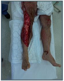
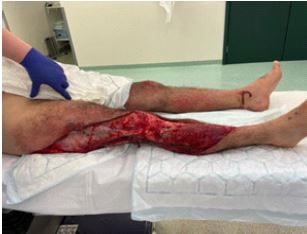
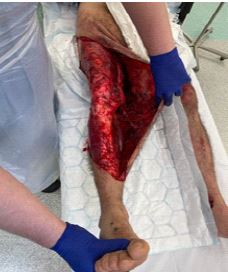
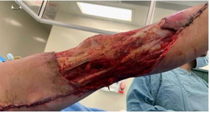
A 45-year-old male presented to the Emergency department following a road cyclist vs Heavy Goods Vehicle collision, having been trapped beneath the wheel of the vehicle for 30 minutes prior to extrication. On initial assessment, an isolated extensive soft tissue injury to the right leg was identified with a significant degloving extending from the medial right ankle to the proximal thigh and buttock (Figures 1-3). The patient had no neurovascular compromise, with a good range of movement of the right knee and ankle. The patient was stabilised in the emergency department, wounds were temporarily dressed, and the patient admitted for combined ortho-plastic management.
Investigations
A trauma CT was performed on arrival to the emergency department which showed a minimally displaced fracture of the proximal right fibula with extensive subcutaneous emphysema and significant soft tissue loss anterior laterally involving the right proximal and distal lower limb. The gas locules extended in to the intermuscular plane both proximally and distally. There were also anterior-lateral fractures of the left 4th and 5th ribs noted with no associated contusion or haemothorax / pneumothorax.
A CT angiogram of the lower limb was subsequently undertaken which showed patency of all iliac and femoral vessels with 3-vessel run-off below the knee.
Treatment
The patient was initially taken to theatre in under 6 hours, undergoing wound debridement and identifying significant contamination with multi-planar degloving injury to the right thigh and lower leg; circumferential at the knee.
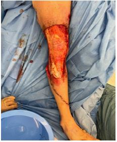
The patient underwent serial debridement’s allowing for demarcation of non-viable tissue, resulting in a 4% Total Body Surface Area (TBSA) wound (Figures 4 & 5). The vastus lateralis had been avulsed from its femoral insertion, leaving tensor fascia lata, semitendinosus and semimembranosus tendons exposed. This injury can be therefore classed as Type 4 multiplanar degloving due to the avulsion of muscle.
This was a wound requiring complex reconstruction. Free tissue transfer was considered at this point, however due the significant progression of tissue necrosis between debridement’s, and risk of further tissue loss, reconstruction with Integra®Dermal Regeneration Template with topical negative pressure dressing was performed.
Over the following 2 weeks the patient returned to theatre a further 3 times for wound inspection and dressing changes, with satisfactory take of the Integra®, until no further debridement was required. The patient was discharged 3 weeks post injury and readmitted 10 days later for second stage reconstruction with meshed split skin grafts, but also further debridement of a necrotic eschar which had developed. The grafts were initially checked 7 days postoperatively, with 100% take. The patient continues to be followed up as an outpatient.

Outcome
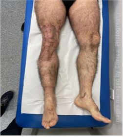
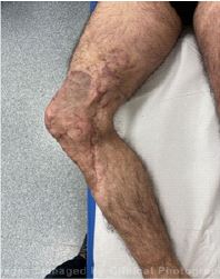
The patient was seen regularly for dressing changes as an outpatient. Small areas of over granulation tissue were treated with a combination of silver dressings and silver nitrate cautery. The patient has had no development of lymphedema post operatively and is able to mobilise independently (Figures 6-8).
Discussion/Conclusion
Degloving injuries are defined as the avulsion of soft tissue whereby an extensive portion of the skin and subcutaneous tissue are avulsed from the underlying fascia, muscles, or bone [3]. These injuries occur as a result of shearing forces being applied to the tissues and in the civilian population are most commonly seen as a result of high energy road traffic accidents [3]. The most common anatomical locations of degloving injuries are the extremities, particularly the lower limb, and truncal areas [4]. However, due to the traumatic nature, many of these injuries lead to decreased vascularity of the wound bed, meaning reconstruction is often challenging [5].
Degloving injuries were first classified in 1999 by Arnezand expanded upon by Khan et al in 2010. Injuries were classified based on the extent and level at which the injury occurs, shown below [2]:
Pattern 1: limited degloving with abrasion / avulsion
Pattern 2: Non circumferential degloving
Pattern 3: Circumferential single plane degloving
Pattern 4: Circumferential multiple plane degloving
Khan et al concluded that all but Pattern 1 injuries required complex reconstruction due to the likely progressive tissue loss as a result of the injuries, and that the use of staged reconstruction in the most severe cases of degloving (Pattern 4) was advisedKhan et al concluded that all but Pattern 1 injuries required complex reconstruction due to the likely progressive tissue loss as a result of the injuries, and that the use of staged reconstruction in the most severe cases of degloving (Pattern 4) was advised due to the development of delayed necrosis. As a result, the patient would require serial debridement’sin order to allow for continued assessment and excision of non-viable tissue prior to definitive reconstruction.
Reconstructive surgeons utilise the reconstructive ladder for techniques to aid in decision making, spanning from healing by secondary intention, to free tissue transfer. Often, multiplanar traumatic injuries may require a combination of techniques depending on which structures require coverage, with exposure of bone and tendon not providing a vascularised wound bed to which a split skin graft would adhere and take requiring a more complex alternative.
Two methods of reconstruction in particular are described in literature. Free tissue transfer, such as the latissimus dorsi or anterolateral thigh flap, provides well vascularised, pliable tissue; however, the extent of the injury can make successful anastomosis away from the zone of trauma challenging. Alternatively, defatting and subsequent replacement of the avulsed skin to the underlying wound bed, as a full thickness skin graft from the degloved skin has been reported [6].
Previously, delayed reconstruction in the context of trauma posed the risk of significant wound infection, however, with the introduction of topical negative pressure dressings and dermal matrix substitutes, such as Integra®, delayed reconstruction has become a viable and commonly used reconstructive option [7].
Integra® is a bilaminar dermal regeneration template; the first layer is composed of a porous, complex matrix of cross-linked bovine tendon collagen and shark glycosaminoglycans which allows for neovascularisation, with a silicone outer layer to act as a barrier to infection [7]. Blood vessels, fibroblasts and myoblasts migrate into the matrix allowing for the formation of the neodermis, whilst the impermeable silicon layer prevents fluid lass and closes the wound [8]. After 4 weeks the matrix is completely replaced by the neodermis [8]. Integra® was initially developed for burn wound coverage but has since been described for use in a variety of wounds such as burns scar revision, diabetic foot ulcers and extremity wounds particularly where coverage of bones, tendons or joints is required [8].
Literature shows that Integra® can provide sufficient coverage of poorly vascularised structures which otherwise would not be suitable for skin grafting. In wounds with exposed tendon or bone, vascularised paratenon or periosteum would have been required for successful skin grafting. In trauma cases however, this is not always present and thus Integra® overcomes these limitations [8]. In this case Integra® was chosen due to the large area of tissue coverage required, exposed bone and the poorly vascularised wound bed, which otherwise would have been unsuitable for skin graft application.
Once dermal regeneration is sufficient, typically taking 3-4 weeks, the silicon layer is removed, and a split-thickness skin graft applied. Reconstruction with Integra® provides a vascularised wound bed suitable for supporting skin graft adherence and healing, as opposed to the relatively avascular exposed bone or tendon. This method holds a number of advantages over other reconstructive techniques such as the large defect created from free tissue harvest. The use of topical negative pressure dressings can be utilised to maintain a sterile environment and reduce shear following skin grafting. It requires regular monitoring for signs of infection with early intervention to prevent destruction of the matrix.
This case highlights the complexity of degloving injuries, demonstrating the need for caution when planning timely reconstruction, and the use of Integra® as a viable option for managing these defects, often with exposure of underlying structures.
Acknowledgement’s: Nill.
References
- Arnez Z, Tyler M, Khan U. Describing severe limb trauma. British Journal of Plastic Surgery. 1999; 52: 280-285.
- Arnez Z, Khan U, Tyler M. Classification of soft-tissue degloving in limb trauma. Journal of Plastic, Reconstructive & Aesthetic Surgery. 2010; 63: 1865-1869.
- Lekuya H, Alenyo R, Kajja I, Bangirana A, Mbiine R, et al. Degloving injuries with versus without underlying fracture in a sub-Saharan African tertiary hospital: A prospective observational study. Journal of Orthopaedic Surgery and Research. 2018; 13.
- Wójcicki P, Wojtkiewicz W, Drozdowski P. Severe lower extremities degloving injuries - medical problems and treatment results. Polish Journal of Surgery. 2011; 83.
- Graham G, Helmer S, Haan J, Khandelwal A. The Use of Integra® Dermal Regeneration Template in the Reconstruction of Traumatic Degloving Injuries. Journal of Burn Care & Research. 2013; 34: 261-266.
- Pilanci O, Akoz Saydam F, Basaran K, Datli A, Guven E, et al. Management of the soft tissue extremity degloving injuries with the full-thickness grafts obtained from the avulsed flap. Turkish Journal of Trauma and Emergency Surgery. 2013; 19: 516-520.
- Attia A, Elmenoufy T, Atta T, Harfoush A, Tarek S, et al. Combination of Negative Pressure Wound Therapy (NPWT) and Integra Dermal Regeneration Template (IDRT) in the lower extremity wound; Our experience with 4 cases. JPRAS Open. 2020; 24: 32-39.
- Chang D, Louis M, Gimenez A, Reece E. The Basics of Integra Dermal Regeneration Template and its Expanding Clinical Applications. Seminars in Plastic Surgery. 2019; 33: 185-189.