
Journal of Clinical Images and Medical Case Reports
ISSN 2766-7820
Case Report - Open Access, Volume 3
A case of Raynaud’s phenomenon in Arizona, USA
Merhavy ZI1, 2*; LaSon DS1,2; Varkey TC2,3
1Ross University School of Medicine; Bridgetown, Barbados, USA.
2Dell Medical School at the University of Texas at Austin; Austin, TX, USA.
3Colangelo College of Business at Grand Canyon University; Phoenix, AZ, USA.
*Corresponding Author : Merhavy ZI
Ross University School of Medicine; Bridgetown, Barbados, USA.
Email: ZackMerhavy@gmail.com
Received : Jun 20, 2022
Accepted : Jul 15, 2022
Published : Jul 22, 2022
Archived : www.jcimcr.org
Copyright : © Merhavy ZI (2022).
Abstract
This case presentation assesses one example of a patient suffering from Raynaud’s phenomenon without having previously sought out medical intervention. This study focuses on the patient’s perspective in hopes of providing insight to future providers in handling patient interactions for those suffering with the same disease. As little about the disease is widely known, this study will address the etiology, pathophysiology, diagnostic tests, current and experimental treatment options, and known genetic components of Raynaud’s disease. The goal of the authorial team is to bring attention to the patient’s perspective to increase rapport, treatment compliance, and increase provider understanding of the language they use to bring up symptomology. This case also looks to help primary care physicians better characterize the patient’s disease state. It is the hope of this research team that available treatments and therapies will become more widely known and understood for the purpose of healthcare professionals to incorporate them into their potential appointments with patients suffering from these conditions.
Keywords: Raynaud; Raynaud’s phenomenon; Raynaud’s disease.
Citation: Merhavy ZI, LaSon DS, Varkey TC. A case of Raynaud’s phenomenon in Arizona, USA. J Clin Images Med Case Rep. 2022; 3(7): 1965.
Background
The patient is a 24-year-old male, living in south Phoenix, Arizona, USA. He has been suffering with Raynaud’s for roughly 8 years at the time this case study was written. The patient initially presented with his chief complaint of hand color changes and painful stimuli when exposed to the cold. He states that two to three of the digits of the hands bilaterally will become pale to white with any minor stimuli of cold exposure and that this leads to increased pain sensation when his hands rewarm. He states the symptoms developed roughly around sophomore year of high school and that they were initially only right sided, but have now become bilateral. He endorses that stress and negative emotional mindsets may cause the phenomena to occur at higher temperatures or become worse, sometimes with him experiencing symptoms up to the mid 70-degree Fahrenheit (~24-degree Celsius) range.
The patient stated that his mother suffered with similar symptoms, but could not remember the name of the disorder, only stating that, “her hands would start to get ‘frostbitten’ very easily, and the first symptom was her hands turning white”. The patient stated that when his mother sought medical attention for her condition, the physician did not know anything about the condition, telling her to “dress intelligently for cold temperatures”. Remembering this experience, the patient had not previously sought out medical attention for himself for the assumption that he would simply be told the same thing.
The patient noted that in addition to the spread and decreasing temperatures of the symptoms in his hands, the onset of symptoms is beginning to occur at faster rates. The patient stated “…because of this, I have to take frequent trips inside to run my hands under warm water so I can avoid any possibility of nerve damage”. He continues to state “I like to go snowboarding, which unfortunately means expensive heated gloves and hand warmers are a must. One time I forgot to bring hand warmers on the mountain and the battery in one of my gloves stopped working after I’d gotten a bunch of snow inside it. After an hour, one finger was turning from white to purple and I had to return to the car for fear of suffering permanent nerve damage. My symptomatic episodes are always confined to my hands and will typically last indefinitely, until I make a real effort to warm up my hands. Just going inside, unless it’s really hot, won’t help most of the time. The fastest method is to run my fingers under warm water which can stop symptoms within a minute or two. The next best option is to hold my fingers in front of the hot air vents in my car, and this can take about 3-5 minutes. If that’s not possible, then I can hold my hands between my armpits, or inside my sleeves, and this can take anywhere from 5-20 minutes. One caveat to all of this is that once symptoms have started the first time, it’s almost like my hands are primed and the issue will return quickly if I don’t stay inside.”
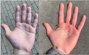
Physical examination of the patient demonstrated hands that would quickly change to white, then with some minor time, the skin would become bluer in coloration. With some rewarming, the patient’s hands would become ruddy in coloration and then return to the patient’s baseline. Upon further investigation, the patient’s pain scale is stated to be at a 2/10 most of the time, stating “this is what makes it scary since sometimes I don’t even notice my fingers have been without blood for a long time”.
Differential diagnosis
Based on the patient’s constellation of symptoms, his family history, and his physical examination the team found that this was most likely a classic case of Raynaud’s syndrome. However, because of the age of the patient, his male biological sex, and the severity of the disease, he was recommended to see an outpatient rheumatologist for further diagnostic work up and treatment. Raynaud’s phenomena can be secondary to a number of disorders and at the time of writing, the patient was working on getting in to see outpatient physicians for treatment of his condition.
The differential for the underlying cause of this particular case includes autoimmune diseases such as Lupus or Scleroderma, medication side effects, especially from drugs that affect smooth muscle contractions (i.e., beta blockers or stimulants), smoking or other allergen side effects, and mechanical injuries to the affected extremities [3,7,8]. Lesser mimics of the disease include Carpal Tunnel Syndrome, minor mechanical injuries from repetitive motions, and increased plaque burden from peripheral arterial disease [3,7,8]. Raynaud’s phenomenon can be secondary to several disorders, and these should be addressed before starting treatment.
End diagnosis and treatment plan
Based on the constellation of symptoms, the patient was diagnosed with Raynaud’s phenomenon and was given instruction and referral paperwork to meet with an outpatient Rheumatologist. As part of the American healthcare system at the time of writing, the patient was working on making an appointment to see an outpatient Rheumatologist for treatment of his condition. He was advised to avoid situations which would impact his digits that may lead to severe nerve damage and was encouraged to dress warmly until the baseline problem was discovered and addressed. As the work-up was still in process, no medications were started at that time.
Summary and etiology
Raynaud’s phenomenon is now a well-established diagnosis, first described in 1862, in a doctoral thesis by Dr. Auguste Gabriel Maurice Raynaud, which consists of a contraction of the smooth muscles of the arteries leading to a decreased perfusion of the capillary beds of downstream tissues. This particular disease affects around five percent (5%) of the global population and is often associated with autoimmune disorders including hypo- or hyperthyroidism, hypo- or hyperparathyroidism, Multiple Sclerosis, and other such disorders [1-5]. The most common location for Raynaud’s phenomenon symptomatology is that of the extremities, particularly the fingers and toes. While more rare, patients with severe disease that need more urgent work describe experiencing symptoms on the tip of the nose, and the ear lobes [1,2,4,5]. Nevertheless, while not often discussed in medical schools, the actual action of this on the smooth muscles of the arteries can be seen all throughout the body and can play a pivotal role in acute care medicine, leading to physicians placing arterial lines in large bore vessels in these patients for fear of auto-amputation [6-8].
The vasoconstriction of the cutaneous arterioles in the extremities and skin is a normal and physiological process which leads to the redirection of the circulation from the surface level capillaries to that of deeper tissues and internal organs. This as a mechanism, teleologically, promotes the conservation of energy by preventing the loss of heat energy from the body and is mediated by the release of norepinephrine, an adrenergic hormone, by the sympathetic nervous system [4-8]. Nevertheless, Raynaud’s phenomenon, a pathological condition, whether primary or secondary, occurs when there is an exaggeration of this normal response in the extremity’s vascular system [1,2,9,10].
Primary Raynaud’s is the disorder itself without a secondary cause [1-5]. Like that of essential or primary hypertension, the diagnosis of Primary Raynaud’s cannot be given until secondary causes, such as autoimmune disorders are ruled out [1-5]. Secondary Raynaud’s occurs when the symptoms of the disorder are caused by another underlying disorder [1-5]. Unlike Primary Raynaud’s, because of the underlying disease state, Secondary Raynaud’s disease often has more severe features and can more quickly progress, leading to soft tissue and/or nerve damage in elevated temperatures as was described in the patient’s case report [1-5].
Pathophysiology
The major biopatho physiological mechanism of action for this disease is directed through the Alpha 2C adrenergic receptor that is found on the vascular smooth muscle cells [3,11]. When these cells are stimulated by the sympathetic nervous system’s release of catecholamines, they increase the amount of calcium ions that enter the smooth muscle cells leading to contraction of the muscle cell itself [12,13]. Normally, on the outside of the cells, there are Alpha 2A and Alpha 2B receptors which induce vasoconstriction and Beta 2A receptors which induce vasodilation [14,16]. Nevertheless, when induced by stress conditions, including cold or other environmental factors, the trans-Golgi receptor, Alpha 2C, is translocated to the cell membrane. When this occurs, the ability of the smooth muscle cells to respond to catecholamines increases exponentially [14,16].
Mechanism of cold-induced mobilization of Alpha 2C adrenergic receptors
During a sudden decrease in temperature, the cutaneous arteriole smooth muscle cells sense this decrease due to changes in the energetics of the breakdown of food molecules [17]. This then causes the mitochondria to release Reactive Oxygen Species (ROS), which then proceeds to activate the Rho/ROCK pathway leading to the rearrangement and reconfiguration of the cytoskeleton [17]. This rearrangement of F-actin and filamin-2 proteins of the cytoskeleton allow for the cell to mobilize the α2C-AR from the endoplasmic reticulum/Golgi to the cell surface [17]. This whole process leads to an increase in the sensitivity of that particular smooth muscle cell, and as a sum total, the entire cutaneous arteriole [17,18]. Other mechanisms by which this process is mediated include the inhibition of gene transcription for the α2C-AR by cyclic AMP’s effect on protein kinase A and the increase in production of α2C-AR by cyclic AMP acting on the exchange protein activated by cyclic AMP (EPAC) and the GTP-binding protein Rap1 [17-18]. The gross exaggeration of these normal biochemical processes and the resulting pathological pain, autoamputation, and/or nerve damage is what is known as Raynaud’s phenomenon or Raynaud’s disease [19].
Tests and diagnosis
In the primary work-up of Raynaud’s disease, similar to other major concerns of medicine is making sure that all the potential causes of the disease are considered. The authors include the major screening tests for the work-up of a patient with Raynaud’s to rule in or out the major baseline etiologies such as the different autoimmune diseases listed above in the Differential section. The main goal of any clinician is to consider the likelihood of the different disease states based on the patient’s history and physical examination, including iatrogenic causes, and then only order those tests which will change the management of the patient [41,42].
Antinuclear Antibody (ANA) test
Antinuclear antibodies are specified antibodies that are found in the blood, programmed to attack the nucleus of the cells within the body’s tissues [20,21]. By performing an ANA test, a positive result can assist healthcare professionals in identifying an autoimmune disorder (i.e., Lupus, Sjogren’s syndrome, Raynaud’s, Scleroderma, etc.) [21]. Although a positive result from an ANA test does not specifically signify Raynaud’s, it may light the way for healthcare professionals to explore further tests to identify the individual’s specific disease. Most autoimmune diseases that occur with secondary Raynaud’s are ANA positive, but many patients with a positive ANA are typically healthy and will remain so [21].
Erythrocyte Sedimentation Rate (ESR)
An Erythrocyte Sedimentation Rate, or ESR, is a type of blood test that measures how quickly red blood cells, or erythrocytes, settle at the bottom of a test tube that contains a blood sample [22]. Normally, red blood cells settle relatively slowly, and thus, a faster-than-normal rate may be caused by inflammation in the body, potentially indicating a sign of a chronic disease, an immune disorder, or other medical condition [22]. An ESR test can help determine if a patient has a condition that causes chronic inflammation such as forms of arthritis, vasculitis, or inflammatory bowel disease [22]. Although an abnormal range from an ESR test does not specifically signify Raynaud’s, it can be used to help a healthcare professional order further tests to identify the individual’s specific disease.
C - Reactive Protein (CRP) Test
A C-Reactive Protein test, or CRP, is often used in conjunction with ESR to indicate a probable chronic inflammatory condition [22,23]. Similarly to ESR, a high CSR value may be a sign of a serious infection or inflammation [22,23]. Both ESR and CRP lack specificity, and thus, should still be used in combination with medical history and a physical exam for diagnosis and monitoring [22]. As many physiological factors such as non-infectious conditions or a resolve of inflammation can contribute to a high ESR and a low CRP or vice versa, it can be incredibly beneficial to a healthcare provider to order both tests [22].
Urinalysis
A urinalysis, or urine dipstick test, can be an invaluable tool for a healthcare provider as it is a cheap and quick method of assisting in a diagnosis [24,25]. A urine dipstick test is a way in which many genitourinary and systemic conditions can potentially be diagnosed [25]. Similar to a CRP test, a urinalysis can detect proteins produced by the liver in response to inflammation and may ultimately help providers determine similar abnormalities seen in autoimmune disorders [24,25]. As the urine dipstick test cannot directly signify whether a patient has Raynaud’s or not, this tool provides means for ruling out other possible chronic autoimmune disorders such as Lupus or glomerulonephritis [24,25].
Complement levels
Testing for complement levels in a patient’s blood can signify whether the pathologic effect is due to an increased or persistent activation, caused by immune complexes as seen in Lupus [28]. Complement levels also help to identify a decreased function or presence of complement inhibitors as seen in cases of ischemia [28]. Based on the specific rise or fall in complement levels that are seen, healthcare providers can determine if a patient likely has a recurrent infection or an autoimmune disease [26]. Again, as much is still unknown about Raynaud’s this test will only further rule out other possible autoimmune disorders as their specific complement indicators have been identified, whereas Raynaud’s has not [26].
Other tests
There are various other lesser-used tests that healthcare professionals can still utilize in order to diagnose Raynaud’s. Many of these highly effective tests, some newer and some older, include Computed Tomography (CT)-guided percutaneous thoracic sympathetic chain radiofrequency thermocoagulation, testing for Th1- and Th17-related cytokines in venous and arterial blood, infrared thermography, photoacoustic and high-frequency ultrasound imaging, and more [12,29-31].
Table 1: This table contains the different tests utilized in the work up of Raynaud’s disease, what they look for, the benefits of ordering the test, and the draw backs to ordering them.
| Test Name | What does it look for? | Benefits | Drawbacks |
|---|---|---|---|
| Antinuclear Antibody (ANA)) | Specific antibodies found in the blood, programmed to attack the nucleus of the cells within the body’s tissues [20-21]. | Can assist in identifying an autoimmune disorder (i.e., Lupus, Sjogren’s syndrome, Raynaud’s, Scleroderma). Is a screening test [20-21]. | Not specific for any particular disease [20-21] |
| Erythrocyte Sedimentation Rate (ESR) | Measures how quickly red blood cells, or erythrocytes, settle at the bottom of a test tube that contains a blood sample [22] | A faster-than-normal rate may be caused by inflammation in the body, potentially indicating a sign of a chronic disease, an immune disorder, or other medical condition. Is a screening test [22] | Not specific for any particular disease [22]. |
| C-Reactive Protein (CRP) | Levels of C-reactive Protein, an acute phase reactant released by the liver secondary to inflammation [22-23]. | A high CSR value may be a sign of a serious infection or inflammation. Is a screening test [22-23]. | Not specific for any particular disease [22-23]. |
| Urinalysis | Abnormal levels of Glucose, Proteins, Ketones, Red Blood Cells, White Blood Cells, or Bacteria within the Urine [24-25]. | A urinalysis can detect proteins produced by the liver in response to inflammation and may ultimately help providers determine similar abnormalities seen in autoimmune disorders [24-25] | Not specific for any particular disease. |
| Complement Levels | Levels of C3 and C4, the major components of the compliment system, activated by antibodies, lectin, or autoactivation [26-28]. | Can signify whether the pathologic effect is due to an increased or persistent activation of the compliment system, caused by immune complexes as seen in Lupus. Can also help to identify a decreased function or presence of complement inhibitors as seen in cases of ischemia [26-28]. | This test will only further rule out other possible autoimmune disorders as their specific complement indicators have been identified [26-28]. |
Table 2: This table includes the major treatment options, their mechanism of action, benefits, and contraindications to use.
| Treatment or therapy | Mechanism of action | Benefit | Contraindication |
|---|---|---|---|
| Alpha Blockers (i.e. Doxazosin, Prazosin, terazosin,tamsulosin, alfuzosin, silodosin, phenoxybenzamine) | Keeping the norepinephrine from tightening the muscles in the walls of smaller arteries and veins, and thus, the vessels remain open and relaxed, ultimately improving blood flow and lowering blood pressure [32]. | Improved the recovery time of finger temperature after being exposed to cold temperatures [32]. | Should be used with caution in patients with a history of postural hypotension, heart failure, and urinary incontinence [32]. |
| Calcium Channel Blockers (i.e.Amlodipine, Diltiazem, Felodipine, Isradipine, Nicardipine, Nifedipine, Nisoldipine, and Verapamil) | Prevent calcium from entering the cells of the heart and arteries [33]. As calcium causes the heart and arteries to contract stronger, by blocking the calcium, blood vessels are allowed to relax and open [1,33] | increases peripheral blood flow and reduces levels of pain experienced during episodes [33] | Sick sinus syndrome (except in those with an artificial pacemaker), pulmonary congestion, acute myocardial infarction,and severe hypotension [1,33]. |
| Nitrate Rich Beetroot Juice | Increases bioavailability of Nitric oxide [34]. | NO-mediated vasodilation, cutaneous vascular conductance and normalized skin temperature following local cooling, and increase systemic anti-inflammatory status [34]. | Congenital heart disease characterized by ductal dependent systemic blood flow,hypersensitivity, and severe left ventricular dysfunction [34]. |
| Single-Port Thoracoscopic Sympathectomy | Removal of the sympathetic nervous connection to that of the digital extremities [35]. | Decrease in the number and duration of Raynaud’s attacks [35]. Useful for those with treatment resistant Raynaud’s phenomenon [35]. | No absolute contraindications to the surgical procedure, however, this particular procedure is still under investigation and will require further evidence before it is considered a quality long-term solution for Raynaud’s phenomenon [35]. |
| Sulodexide (~80% heparan sulfate and ~20% dermatan sulfate) | Components of the arterial wall, or basal membrane, and extracellular matrix, which ultimately influences the function and activity of heparin-binding proteins decreasing the likelihood of clot formation within constricted blood vessels and capillaries of the extremities [36]. | Significant increase in blood perfusion in the capillary vessels, a reduction in the frequency of pain episodes, and a decrease of pain intensity during episodes [36]. | Diathesis, Hemorrhagic diathesis, and Hypersensitivity (allergy) to any component of sulodexide [36]. |
Treatments and therapies
As the patient referenced, physicians have only commented on proper dress for weather conditions rather than providing a solution, therapy, or treatment options to his mother who suffers from the same condition, ultimately leaving the patient feeling as though seeking treatment would be pointless. Although there is not currently an abundance of treatments or therapies known for Raynaud’s, it is still important to identify ones that do exist.
Alpha blockers
Alpha blockers are known for being one of the two most widely used treatment options with a high rate of success in many patients [1]. Alpha blockers work by keeping the norepinephrine from tightening the muscles in the walls of smaller arteries and veins, and thus, the vessels remain open and relaxed, ultimately improving blood flow and lowering blood pressure [32]. One study discovered that use of alpha blockers, with or without a combination of a beta blocker, improved the recovery of finger temperature after being exposed to cold temperatures [32].
Calcium channel blockers
Calcium channel blockers are the second of the two most widely used treatment options with a high rate of success in many patients, often serving as the first choice of treatment for affected patients [1]. Calcium channel blockers ultimately work similarly to alpha blockers, only by preventing calcium from entering the cells of the heart and arteries [33]. As calcium causes the heart and arteries to contract stronger, by blocking the calcium, blood vessels are allowed to relax and open [33]. Use of calcium channel blockers in patients with Raynaud’s, it has been observed that this medication increases peripheral blood flow and reduces levels of pain experienced during episodes [33].
Beetroot juice
As it is known that Raynaud’s phenomenon is characterized by recurrent transient peripheral vasospasm and lower Nitric Oxide (NO) bioavailability in the cold, a research team investigated the effect of nitrate-rich beetroot juice supplementation on NO-mediated vasodilation, cutaneous vascular conductance and skin temperature following local cooling, and systemic anti-inflammatory status [34]. After the participants drank the beetroot juice every day for 2 weeks, the research team discovered that each participant saw an improvement in thumb blood flow, improved endothelial function and anti-inflammatory status, as well as a reduction in BP [34].
Single-port thoracoscopic sympathectomy
Single-port thoracoscopic sympathectomy is a novel, miniminimally invasive endoscopic technique used in patients with treatment-resistant Raynaud’s [35]. In another study, this was performed unilaterally on the left side in eight patients with Raynaud’s, and after surgery on the left hand only, a unilateral improvement in perfusion was observed in the left hand compared with the right hand [35]. Additionally, the number and duration of attacks in the left hand had decreased over a 2-week period as compared with the right hand with no serious adverse events occurring in a follow-up period of 10 months [35].
Sulodexide
Sulodexide is a mixture of glycosaminoglycans (~80% heparan sulfate and ~20% dermatan sulfate), which are components of the arterial wall, or basal membrane, and extracellular matrix, and ultimately influences the function and activity of heparin-binding proteins [36]. Although calcium antagonists, angiotensin-converting enzyme inhibitors, and α1-blockers are currently the most common medications in the pharmacotherapy of Raynaud’s, one study found that sulodexide improved capillary blood flow in patients with Raynaud’s [36]. A significant increase in blood perfusion in the capillary vessels was observed as well as a reduction in the frequency of pain episodes and a decrease of pain intensity during episodes [36].
Genetics
Variations in genes may be involved in the process of excessive constriction of small blood vessels in response to cold or stress; however, the exact connection between these gene variations and the abnormal blood vessel response that occurs in this disorder is currently unknown [37,43]. The main reasoning for this unknown connection is due to the lack of robust biomarkers needed to predict vascular outcomes or responses to therapy in patients with Raynaud’s [38]. It has been observed that certain cases of Raynaud’s may be familial, but the specific inheritance pattern is still unknown [37,43]. Some studies suggest that roughly 30% of individuals with a first-degree relative, such as a parent, sibling, or child, who has primary Raynaud’s phenomenon may also have the condition as well [37]. Although the exact genetic component has not been clarified, one study suggests that hyperhomocysteinemia resulting from methylenetetrahydrofolate reductase, or MTHFR, gene mutation may have a role in Raynaud’s etiology [39]. Most individuals observed with this MTHFR gene mutation and hyperhomocysteinemia were, more frequently than not, diagnosed with Raynaud’s phenomenon, making up an estimated 3-20% of the global population [39].
In cases of secondary Raynaud’s, this exaggerated vasoconstriction could be caused by autoimmune diseases; hypothyroidism; cancers of the blood, bone marrow, or immune system; disease processes that cause obstruction of blood vessels; exposure to certain medicines or chemicals; cigarette smoking; injury or trauma; prolonged repetitive motions such as typing; long-term use of vibrating tools; and more [37,40].
Social impact
When asked about the impact his condition has on his social life, the patient attributed the “uncertainty” of when the symptoms will arise as the leading cause of difficulty to plan around his condition; having to always dress warm just in case, always have hand warmers available, and to make sure he has heated gloves. He additionally notes that a “negative or passive” mental state plays a large factor as well, naming stress and/or anxiety some of the worst culprits. The patient notes the irony that his condition brings forth these stressors, which in turn, causes the symptoms to get worse, stating “It’s a constant source of stress when I’m anywhere the temperature gets even a little chilly”.
He continues to comment by stating “The symptoms mostly impact my social life. If I’m out to dinner or a bar with friends who want to sit outside, I don’t want to be the one to have everyone sit inside since I have an obscure condition that nobody understands. Especially when I’m talking to someone when it’s a little chilly outside, I find that sometimes I can’t focus on the conversations happening around me because I’m so focused on keeping my hands warm.
Unfortunately, most of the times I spend prolonged periods outside are social outings with friends and peers. For someone who is prone to stress and anxiety to begin with, it can be very problematic and results in a sort of feedback loop. The fear and anxiety at the thought of my fingers losing blood flow causes the symptoms in the first place, and it becomes a kind of self-fulfilling prophecy. Suddenly, I can no longer enjoy my relaxing patio dinner, or fun camping trip, or whatever it may be. All I can think about is how I need to warm up my fingers or risk nerve damage, which is conjoined by the anxiety and stress from the knowledge that, yet again, I won’t be able to fully enjoy a social event due to my condition”.
Provider considerations
Based on the information presented thus far in this particular article, it is key that some recommendations for clinical practice be considered. First and foremost is the patient-centered care that must occur. For this aspect, the provider should ask the patient about the temperature at which the patient is experiencing the symptoms, how much this bothers them, provide them all alternatives of treatment/therapeutic options, and then ask what option that they are desirous to pursue. Through ensuring that the patient is given all the pertinent information and is the one making the decision, the patient’s autonomy and their wishes are respected. Secondarily, the provider should provide an explanation of the condition at the patient’s level to ensure that they understand the condition and what it means. Finally, the provider needs to ensure that the patient is able to explain what the condition is and how they are planning on treating it using a method like that of the teach back method, where the patient provides a detailed explanation of the disease process and their medications/lifestyle modifications to the provider where any misunderstanding can be corrected. Through placing the patient and their care first and foremost, the provider not only improves their relationship with the patients, but they also increase the likelihood that the patient will follow through with the shared treatment plan - increasing the likelihood of positive outcomes for the patient.
References
- Herrick AL. Pathogenesis of Raynaud’s phenomenon. Rheumatology, 2005; 44: 587-596.
- Lis Święty A. Recent advances in the workup and management of Raynaud phenomenon. Polish Archives of Internal Medicine, 2019; 129: 798-808.
- Haque A, Hughes M. Raynaud’s phenomenon. Clinical Medicine. 2020; 20: 580-587.
- National Heart, Lung, and Blood Institute. Raynaud’s. Retrieved from https://www.nhlbi.nih.gov/health-topics/raynauds. 2021.
- National Institute of Arthritis and Musculoskeletal and Skin Diseases. Raynaud’s phenomenon. Retrieved from https://www.niams.nih.gov/health-topics/raynauds-phenomenon. 2016.
- Charkoudian N, Stachenfeld N. Sex hormone effects on autonomic mechanisms of thermoregulation in humans. Auton, Neurosci. 2016; 196: 75–80.
- Vanhoutte PM. Physical factors and regulation of vascular smooth muscle function, in Handbook of Physiology, eds Bohr DF, Somlyo AP, Sparks HV. (Washington, DC: The American Physiological Society), 1980; 443–474.
- Wigley FM, Flavahan NA. Raynaud’s phenomenon. N. Engl. J. Med. 2016; 375: 556–565.
- Herrick AL. The pathogenesis, diagnosis and treatment of Raynaud phenomenon. Nat. Rev. Rheumatol. 2012; 8: 469–479.
- Cordeiro RA, de Andrade RM. Raynaud’s phenomenon in the occupational context. Revista da Associacao Medica Brasileira. 2019; 65: 1314-1320.
- Nicola S, Fornero M, Fusaro E, Peroni C, Priora M, Rolla G, et al. Th1- and Th17-related cytokines in venous and arterial blood of sclerodermic patients with and without digital ulcers. BioMed Research International, 2019; 2019: 7908793.
- Rimar D, Rimar O, Rosner I, Rozenbaum M, Kaly L, et al. Nailfold video capillaroscopy: Beyond systemic sclerosis. The Israel Medical Association Journal. 2019; 21.
- Dinsdale G, Wilkinson S, Wilkinson J, Moore TL, Manning JB, Berks M, et al. State-of-the-art technologies provide new insights linking skin and blood vessel abnormalities in SSc-related disorders. Microvascular Research. 2020; 130: 104006.
- Di Benedetto P, Ruscitti P, Liakouli V, Cipriani P, Giacomelli R, et al. The vessels contribute to fibrosis in systemic sclerosis. The Israel Medical Association Journal. 2019; 21: 471-474.
- Soloway AM, DePace NL, Colombo J, Soloway S, et al. Cyanosis with dysautonomia mimics Raynaud disease. Journal of Clinical Rheumatology: Practical Reports on Rheumatic & Musculoskeletal Diseases. 2020; 26:254-255.
- Nagaraja V, Matucci Cerinic M, Furst DE, Kuwana M, Allanore Y, et al. Current and future outlook on disease modification and defining low disease activity in systemic sclerosis. Arthritis & Rheumatology. 2020; 72: 1049-1058.
- Fardoun MM, Nassif J, Issa K, Baydoun E, Eid AH. Raynaud’s Phenomenon: A Brief Review of the Underlying Mechanisms. Frontiers in pharmacology. 2016; 7: 438.
- Herrick AL. The pathogenesis, diagnosis and treatment of Raynaud phenomenon. Nat Rev Rheumatol. 2012; 8: 469-479.
- Eid AH, Chotani MA, Mitra S, Miller TJ, Flavahan NA, et al. Cyclic AMP acts through Rap1 and JNK signaling to increase expression of cutaneous smooth muscle alpha2C-adrenoceptors. Am J Physiol Heart Circ Physiol. 2008; 295: H266-H272.
- Mayo Clinic. Raynaud’s disease. Retrieved fromhttps://www.mayoclinic.org/diseases-conditions/raynauds-disease/symptoms-causes/syc-20363571?p=1 2021.
- O’Sullivan M, McLean Tooke A, Loh RK. Antinuclear antibody test. Australian Family Physician. 2013; 42: 718-721.
- Bray C, Bell LN, Liang H, Haykal R, Kaiksow F, Mazza JJ, et al. Erythrocyte Sedimentation Rate and C-Reactive Protein measurements and their relevance in clinical medicine. Wisconsin Medical Journal. 2016; 115: 317-321.
- Ahrazoglu M, Moinzadeh P, Hunzelmann N. Differenzialdiagnosen des Raynaud-Syndroms [Differential diagnoses of Raynaud’s phenomenon]. Deutsche MedizinischeWochenschrift. 2014; 139: 1064-1069.
- Pauling JD, Hughes M, Pope JE. Raynaud’s phenomenon-au update on diagnosis, classification and management. Clinical Rheumatology, 2019; 38: 3317-3330.
- Sharp VJ, Lee DK, Askeland EJ. Urinalysis: Case presentations for the primary care physician. American Family Physician. 2014; 90: 542-547.
- Kirschfink M, Mollnes TE. Modern complement analysis. Clinical and Diagnostic Laboratory Immunology. 2003; 10: 982-989.
- Bournia VK, Kottas K, Panopoulos S, Konstantonis G, Iliopoulos A, et al. Differential performance of nailfold video capillaroscopic parameters in the diagnosis and prognosis of systemic sclerosis. Clinical and Experimental Rheumatology. 2020; 125: 29-39.
- Nilsson B, Ekdahl KN, et al. Complement diagnostics: Concepts, indications, and practical guidelines. Clinical & Developmental Immunology. 2012; 2012.
- Martini G, Cappella M, Culpo R, Vittadello F, Sprocati M, et al. Infrared thermography in children: A reliable tool for differential diagnosis of peripheral microvascular dysfunction and Raynaud’s phenomenon?. Pediatric Rheumatology Online Journal. 2019; 17: 68.
- Huang H, Qiu W, Chen Q, Sun K, Huang B, Computed Tomography (CT)-guided percutaneous thoracic sympathetic chain radiofrequency thermocoagulation for Raynaud disease. Medical Science Monitor: International Medical Journal of Experimental and Clinical Research. 2019; 25: 7391-7395.
- Daoudi K, Kersten BE, van den Ende CHM, van den Hoogen FHJ, Vonk MC. Photoacoustic and high-frequency ultrasound imaging of systemic sclerosis patients. Arthritis Research & Therapy. 2021; 23: 22.
- Cleophas TJM, van Lier HJJ, Fennis JFM, van’t Laar A, et al. Treatment of Raynaud’s Syndrome with Adrenergic Alpha-Blockade with or without Beta-Blockade. Angiology. 1984; 35: 29-37.
- Smith CR, Rodeheffer RJ. Treatment of Raynaud’s phenomenon with calcium channel blockers. The American Journal of Medicine. 1985; 78: 39-42.
- Shepherd AI, Costello JT, Bailey SJ, Bishop N, Wadley AJ, et al. Beet the cold: Beetroot juice supplementation improves peripheral blood flow, endothelial function, and anti-inflammatory status in individuals with Raynaud’s phenomenon. Journal of Applied Physiology. 2019; 127: 1478-1490.
- van Roon AM, Kuijpers M, van de Zande SC, Abdulle AE, van Roon AM, et al. Treatment of resistant Raynaud’s phenomenon with single-port thoracoscopic sympathicotomy: A novel minimally invasive endoscopic technique. Rheumatology. 2020; 59: 1021-1025.
- Madycki G, Obidzińska Trościanko J, Juszyński M, Zgliczyński W, Glinicki P, et al. Sulodexide improves capillary blood flow and the quality of life in patients with Raynaud syndrome: A pilot study. Polish archives of internal medicine. 2020; 130: 79-81.
- Pistorius MA, Planchon B, Schott JJ, Lemarec H. Aspects héréditaires et génétiques de la maladie de Raynaud [Hereditary and genetic aspects of Raynaud’s disease]. Journal des Maladies Vasculaires. 2006; 31:10-15.
- Mecoli CA, Perin J, Van Eyk JE, Zhu J, Fu Q, Allmon AG, et al. Vascular biomarkers and digital ulceration in systemic sclerosis: Results from a randomized controlled trial of oral treprostinil (DISTOL-1). Clinical Rheumatology. 2020; 39: 1199-1205.
- Yalçin KS, Koşar A. The frequency of Raynaud’s phenomenon in patients with methylenetetrahydrofolate reductase gene mutation and hyperhomocysteinemia. Turkish Journal of Medical Sciences. 2019; 49: 1444-1449.
- Ferri C, Giuggioli D, Guiducci S, Lumetti F, Bajocchi G, Magnani L, et al. Systemic sclerosis Progression INvestiGation (SPRING) Italian registry: Demographic and clinico-serological features of the scleroderma spectrum. Clinical and Experimental Rheumatology. 125: 40-47.
- Novikov D, Cizmic Z, Feng JE, Iorio R, Meftah M, et al. The historical development of value-based care: How we got here. The Journal of Bone & Joint Surgery. 2018; 100: e144.
- Siu VJ, Varkey T, Khan UN, Ding JB, Gandhi S, et al. Lend me a hand: A value-based care case study on pan plexopathy of unknown origin. Cureus. 2021; 13: e20354.
- Cherkas LF, Williams FM, Carter L, Howell K, Black CM, et al. Heritability of Raynaud’s phenomenon and vascular responsiveness to cold: A study of adult female twins. Arthritis Care & Research. 2007; 57: 524-528.