
Journal of Clinical Images and Medical Case Reports
ISSN 2766-7820
Short Report - Open Access, Volume 3
Recrudescent pouch calculi in cutaneous continent diversion
K Sricharan Raj; Padmaraj Hegde*; Mayank Kulshrestha; Naman Aggarwal; GVS Abhishek; BV Reddy
Department of Urology and Renal Transplant, Kasturba Hospital, Manipal, Karnataka, India.
*Corresponding Author : Padmaraj Hegde
Professor, Department of Urology and Renal Transplant, Kasturba Hospital, Manipal, Karnataka, India.
Email: padmaraj.hegde@manipal.edu
Received : Jun 23, 2022
Accepted : Jul 19, 2022
Published : Jul 26, 2022
Archived : www.jcimcr.org
Copyright : © Padmaraj H (2022).
Abstract
Reservoir pouch calculi are the most common complication following continent urinary diversion. The presence of calculi in the neobladder can be silent or provoke flank pain, hematuria, and urinary tract infections. Minimal invasive interventions like endoscopic or percutaneous retrieval in smaller calculi to open surgical approaches in large calculi is some of the treatment modalities. Here we present a 34-year-old patient who is a known case of exstrophy-epispadias complex, underwent bladder closure at one year of age and continent cutaneous urinary diversion (Modified Indiana pouch) 26 years ago. Plain and contrast computerized tomography showed multiple large calculi six in number, inside the pouch. In this case, the calculi were successfully managed through an open surgical approach.
Keywords: Pouch calculi; Modified indiana pouch; Poucholithotomy.
Citation: Raj KS, Hegde P, Kulshrestha M, Aggarwal N, Abhishek GVS, et al. Recrudescent pouch calculi in cutaneous continent diversion. J Clin Images Med Case Rep. 2022; 3(7): 1969.
Introduction
Reservoir pouch calculi are the most common complication following continent urinary diversion. In the continent cutaneous diversion, calculi appear to be more prevalent than orthotopic diversions [1]. The formation of pouch calculi is driven by urinary stasis caused by poor voiding, urinary infection with urea-splitting bacteria, encrustation on a foreign body, mucus, and metabolic acidosis [2]. After Indiana pouch modification, the reported incidence of stones associated with urinary diversion ranged from 11% to 12.9%. Furthermore, within 3-5 years of prompt treatment, these stones have a recurrence rate of 33-63 % [3]. The presence of calculi in the neobladder can be silent or provoke flank pain, hematuria, and urinary tract infections. Minimal invasive interventions like endoscopic or percutaneous retrieval in smaller calculi to open approaches in large calculi are some of the treatment modalities [4]. We present a case of recurrent pouch calculi in post-cutaneous continent diversion and its successful management through an open surgical approach.
Case history
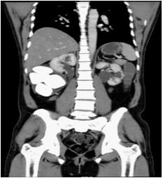
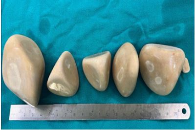
A 34-year-old male presented with fullness in the flank and intermittent left flank pain for two months. Patient is a known case of exstrophy-epispadias complex who underwent bladder closure at one year of age and continent cutaneous urinary diversion (Modified Indiana pouch) 26 years ago. The patient had a history of Poucholithotomy 12 years back for similar complaints. A plain X-ray showed radiopaque shadow in the right lumbar region. Plain and contrast computerized tomography showed multiple large calculi six in number, with the most giant measuring 5 cm × 3.6 cm hyperdense calculi (1035 HU) inside the pouch (Figure 1). Open poucholithotomy was performed through a right-sided paramedian incision, medial to the stoma. Neobladder was identified and was opened along the teniae coli. Complete stone clearance was achieved. Six calculi were removed intact (Figure 2). The neobladder was closed in two layers, and a 24 Fr abdominal drain was placed. Neo-bladder was catheterized with 14 Fr Foleys. The perioperative period went off without a hitch, and the drain was removed on day four after surgery. On the fifth day, the patient was discharged with instructions for regular catheterization and follow-up. After three weeks, the catheter was removed and regular clean intermittent catheterization was resumed. Calcium phosphate, calcium carbonate, and urate were found in the stone.
Discussion
The management of urinary calculi in the neobladder is challenging. However, the therapeutic concepts are essentially unaltered from those used to treat calculi in a regular bladder. Laparoscopic and Endoscopic retrieval, percutaneous removal of calculi have all been recorded in patients with modest stone burden [5]. Endoscopic management using a Mitrofanoff catheterizable conduit is not encouraged since the continence mechanism may be disrupted [6]. For large stone loads, open cystolithotomy is often preferable. Despite the fact that urolithiasis is managed in such cases, recurrence is common. Cohen et al. observed a 63% recurrence rate after a 5-year follow-up period [7]. The reservoir’s constant mucus production creates a nidus for stone recurrence. This emphasises the need of proper reservoir drainage and irrigation in preventing stone recurrence.
Stone recurrence is fostered by persistent mucus production in the reservoir. This emphasises the need of proper reservoir drainage and irrigation in minimizing recurrence of stones [8]. Open poucholithotomy was preferred in our case, as it seemed the best option for managing such an enormous stone burden in the pouch. Regular and efficient reservoir draining, can help to reduce stone recurrence within continent diversion.
References
- Terai A, Ueda T, Kakehi Y, et al. Urinary calculi as a late complication of the Indiana continent urinary diversion: Comparison with the Kock pouch procedure. J Urol. 1996; 155: 66–68.
- Holmes DG, Thrasher JB, Park GY, et al. Long-term complications related to the modified Indiana pouch. Urology. 2002; 60: 603–606.
- Zhong W, Yang B, He F, Wang L, Swami S, et al. Surgical management of urolithiasis in patients after urinary diversion. PLoS One. 2014; 9: e111371.
- Jesse E, Mahdy A. Surgical Management of large pouch stone in continent urinary diversion. Int Urogynecol J. 2018; 29: 165-166.
- Benson MC, Olsson CA. Continent urinary diversion. Urol Clin North Am. 1999; 26: 125-147, ix
- Lam PN, Te CC, Wong C, Kropp BP. Percutaneous cystolithotomy of large urinary-diversion calculi using a combination of laparoscopic and endourologic techniques. J Endourol. 2007; 21: 155-157.
- Cohen TD, Streem SB, Lammert G. Long-term incidence and risks for recurrent stones following contemporary management of upper tract calculi in patients with a urinary diversion. J Urol. 1996; 155: 62-65.
- L’Esperance JO, Sung J, Marguet C, L’Esperance A, Albala DM, et al. The surgical management of stones in patients with urinary diversions. Curr Opin Urol. 2004; 14: 129-134.