
Journal of Clinical Images and Medical Case Reports
ISSN 2766-7820
Case Report - Open Access, Volume 3
Imperforate hymen causing hematometrocolpos and acute urinary retention
Fernando Javier Peralta Palmezano1*; Diana Paola Escobar Serna2,3; Juan Javier Peralta Palmezano2,3
1Department of Gynecology and Obstetrics, Universidad de Antioquia, Medellin, Colombia.
2Department of Pediatrics, Universidad Nacional de Colombia, Bogotá, Colombia.
3HOMI Fundación Hospital Pediátrico la Misericordia, Bogotá, Colombia.
*Corresponding Author : Fernando J Peralta-Palmezano
HOMI Fundación Hospital Pediátrico la Misericordia, Avenida Caracas # 1-65. Bogotá, Colombia.
Phone: (+57) 301 750 4163, Fax: +57-1-309-3313;
Email: fjperaltap@unal.edu.co
ORCID ID: 0000-0002-0427-5998
Received : Jul 04, 2022
Accepted : Jul 26, 2022
Published : Aug 02, 2022
Archived : www.jcimcr.org
Copyright : © Peralta-Palmezano FJ (2022).
Abstract
Introduction: The imperforate hymen is the result of a failure in the canalization of the vaginal plate, with an incidence of 1 in 2000 live births of women. Imperforate hymen can complicate with hematocolpos or hematometrocolpos. Common clinical manifestation is a periodic and long-lasting abdominal pain, enduring months to years before diagnosis.
Case presentation: We present an 11-year and nine-month-old patient, admitted to our emergency department without imperforate hymen typical manifestations. She consulted for a first episode of lower abdominal pain and acute urinary retention. Hematometrocolpos, an imperforate hymen complication, was diagnosed with ultrasonography and magnetic resonance imaging. Genital inspection showed an imperforate, bulging, and bluish hymen at the introitus. Our patient was operated and discharged next day.
Conclusion: Imperforate hymen is easy to diagnose when a complete physical exam is performed. Premenarchal girls with abdominal pain and acute urinary retention, should be suspected imperforate hymen even in a first episode of abdominal pain.
Keywords: Abdominal pain; Adolescent; Case reports; Hematocolpos; Hematometra.
Citation: Peralta-Palmezano FJ, Escobar-Serna DP, Peralta-Palmezano JJ. Imperforate hymen causing hematometrocolpos and acute urinary retention. J Clin Images Med Case Rep. 2022; 3(8): 1980.
Introduction
The development of vaginal plate occurs by combining the Müllerian ducts, the urogenital sinus, and the Wolffian ducts. When there is a failure in the canalization of the vaginal plate, the imperforate hymen is produced [1]. The incidence of imperforate hymen is 1 per 2000 live births of women [2]. It is the most frequent congenital obstructive anomaly of the genital tract and can be associated with other malformations of the genitourinary tract in 20% of cases [3]. 9.3% of patients have a family history of imperforate hymen [3].
It is usually an asymptomatic condition until menarche, when there is accumulation of menstrual blood in the vagina (hematocolpos) or in the vagina and uterus (hematometrocolpos). Complicated imperforate hymen can give rise to a variety of symptoms, the most common being long-lasting, cyclical lower abdominal pain that worsens over time. It can be associated with low back pain, constipation, dysuria, urinary retention, urinary tract infection and kidney failure [3,4]. Acute urinary retention (AUR) is a rare symptom and consists of the inability to empty the bladder voluntarily for more than 12 hours, with a volume of urine greater than expected for age or a distended and palpable bladder [5]. The main causes of AUR in adolescents are: urolithiasis, urinary tract infections, neurological disorders, trauma, constipation, malignancies and medications [5,6]. The imperforate hymen represents only 3.5% of the cases [6]. Our case report is relevant because we present an adolescent with an imperforate hymen, who was admitted for a first episode of abdominal pain and AUR.
Case presentation
With prior informed consent signed by the legal representative and authorized by the patient, we present an 11-year and nine-month-old patient who attends the emergency department of our hospital. Our patient had two days of lower abdominal pain, cramp-like, increasing in intensity, and associated with vomits, dysuria and urine inability for 24 hours, without fever. Also, absence of depositions for four days prior to admission. She had no previous abdominal pain or urinary symptoms. Days before consulting us, our patient was managed with bladder catheterization and discharged in another hospital. Because AUR reappeared, she consults our hospital. Her antecedents were corpus callosum agenesis, chronic constipation, and appendectomy at three years. No important family history. Thelarche at nine years, without menarche, and she was not sexually active.
Our patient was phenotypically normal and had Tanner stage III pubertal development. Initial physical examination evidenced a distended abdomen with a painful suprapubic mass and no peritoneal irritation signs, rectal and genital examination is omitted. With an AUR diagnosis and chronic constipation we initiated analgesic, laxatives, and bladder catheterization, draining 550 ml of clear urine, and improving abdominal distension and pain.
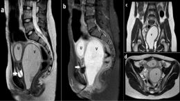
Urinalysis, blood count, blood urea nitrogen, and creatinine were normal. Abdominal ultrasonography showed an 81 x 70 x 113 mm hypoechoic collection, compatible with hematometra, with additional bicorne uterus suspicion, without renal, ureteral o bladder abnormalities. Because of ultrasonographic abnormalities, we performed a contrasting pelvic magnetic resonance imaging, finding normal ovaries and an uterus with usual morphology. Magnetic resonance imaging showed homogeneous material distending endometrial cavity (measured 53x 21 x 37 mm) and vaginal canal (measured 116 x 53 x 56 mm), compatible with hematometrocolpos (Figure 1). Genital inspection showed a normal vulva with an imperforate, bluish, and bulging membrane in the introitus (Figure 2a).
After general anesthesia and antibiotic prophylaxis, the hospital´s pediatric surgeon opened by vertical incision the hymen (hymenotomy), draining 300 ml of dark, non-fetid blood. Then resected hymen membrane edges (hymenectomy) and sutured hymeneal folds with interrupted absorbable suture (vicryl 5-0) (Figure 2b). Post-surgical recovery of our patient had no complications and bladder catheter is removed, verifying spontaneous urination. She is discharged the next day after surgery. Six weeks after surgery she was asymptomatic and with menstruation from the fourth week after surgery, confirming hymeneal permeability.

Discussion
Symptoms secondary to imperforate hymen, in premenarchal girls are caused by menstrual blood efflux inability, resulting in hematocolpos or hematometrocolpos. It manifests mainly with abdominal pain, abdominal and/or pelvic mass, and genital examination shows a tense and bluish membrane in the introitus [7-9]. In the case presented, AUR was the cardinal sign that guided the diagnosis and subsequent studies confirmed hematometrocolpos secondary to imperforate hymen. In the patient, the gynecological examination was omitted until the imaging studies, being a late diagnosis.
In women, urinary retention is rare because of its shorter, straighter urethra with a wider diameter. Hematocolpos causes urinary retention obstructing the urethra by either a narrowing or angulation, as well as causing sacral plexus irritation [9]. Our magnetic resonance imaging (Figure 1a and 1b) showed compression and angulation in the urethra by hematocolpos, supporting the pathophysiological mechanism mentioned previously. Studies with urodynamics show an increase in maximal urethral closure pressure due to an urethra compression by hematocolpos, but these patients can urinate normally whenever they want. Acute urinary retention occurs when urinary volume exceeds maximal urethral closure pressure threshold, which depends on each patient. One way to verify this in an asymptomatic patient is to have her hold urine for prolonged periods of time [9]. Our patient was initially managed with bladder catheterization for AUR in another hospital. After urinary evacuation, she was sent home with no additional management or recommendations in voiding habits. Her AUR reappearance would be secondary to an inadequate voiding habit after discharge.
Images are usually not indicated for a classic presentation of imperforate hymen, which can be clinically diagnosed. Ultrasonography is first choice with rectal visualization being better. Ultrasonography is also indicated in cases where the diagnosis is uncertain, when other utero-vaginal obstructive anomalies are suspected, to detect renal anomalies, in the follow-up of hydronephrosis and endometriosis and the documentation of genital decompression after surgery [8]. Magnetic resonance imaging should be performed not only to confirm diagnosis and evaluate the collection extent, but also to rule out other urogenital tract anatomical variants and avoid unnecessary surgical interventions [11]. It is indicated when there are limitations in the evaluation with ultrasonography. Imperforate hymen repair can be performed at any age, as long as it does not resolve with estrogenization (at 1 to 2 months of age or 1 year after thelarche). In adolescence, estrogenization will improve the healing of the hymenal resection [9]. Definitive management of the imperforate hymen is a hymenotomy, after urinary catheterization, evacuating the accumulated blood. Hymenotomy is performed with a cruciform or T-shaped incision, followed by hymenectomy and suturing of the hymenal ring with absorbable suture to prevent adhesions and bleeding. Puncture and drainage alone, without definitive hymenal resection, should be avoided, as this can cause scarring and ascending infection [9]. Conservative techniques with circular or elliptical hymenotomy, with or without Foley catheter placement [12], are not universally accepted due to the risk of scarring, infections, and recurrence. We performed a vertical hymenotomy with annular hymenectomy. Results after a hymenotomy are usually excellent, however, follow-up is necessary to ensure the absence of recurrence or symptoms such as dyspareunia, dysmenorrhea, abnormal menstruation, persistent urination/defecation, or fertility problems [13].
Chronic constipation causes AUR [5,6]. Our patient had no bowel movements previous days attending the emergency department. We managed her with laxative, without improvement of AUR. After imaging and genital examination findings; we focused a genital tract obstruction as the cause of AUR. Girls with imperforate hymen often do not present with gynecological symptoms and although diagnosis is easy, with a detailed anamnesis and a physical examination, it can be overlooked and lead to unnecessary diagnostic tests, inadequate treatment or delay the specific diagnosis and future complications [14].
It is diagnosed in the neonatal period in only 11% of patients, manifesting with hydrometrocolpos [3]. In an adolescent with abdominal pain, with breast Tanner stage III or IV, without having presented menarche, the diagnosis of imperforate hymen should be assessed [3,10]. Prognosis depends on early detection and treatment and the severity of associated anomalies [15]. Evaluation of the external genitalia should be routine practice in pediatrics [15].
Conclusion
Patients with imperforate hymen complicated with hematometrocolpos consult the emergency department with abdominal pain and acute urinary retention. Imperforate hymen´s typical clinical manifestations are periodic and long-lasting abdominal pain in a premenarchal girl. Our case never had abdominal pain antecedents and because the previous symptoms absence, there was a diagnosis delay. Imperforate hymen should be considered as an etiology when evaluating premenarchal adolescents with low abdominal pain, AUR, and pelvic mass. Imperforated hymen should be suspected in premenarchal girls with a first episode of abdominal pain and acute urinary retention without a clear cause.
Declarations
Ethical approval: Our case report was approved by the Institutional Review Board of the HOMI Fundación Hospital Pediátrico la Misericordia (ethics board approval number: CEI 226-19).
The work described has been carried out in accordance with The Code of Ethics of the World Medical Association (Declaration of Helsinki)
Contributors: All authors contributed to the conception, drafting, review, and revision of the manuscript. All authors read and approved the final version of the paper and take full responsibility for the work.
Financing: No financial support was received in relation to this study.
Conflict of interest: Authors declare that the research was conducted in absence of any commercial or financial relationships that could be construed as a potential conflict of interest.
References
- Luthra, M. & Douglas Stephens, F. Embryogenesis of the hymen and caudal end of the vagina deduced from uterovaginal anomalies Pediatr Surg Int. 1988; 3: 422-425.
- Parazzini F, Cecchetti G. The frequency of imperforate hymen in northern Italy. Int J Epidemiol. 1990; 19(3): 763-4.
- Lee KH, Hong JS, Jung HJ, Jeong HK, Moon SJ, Park WH, et al. Imperforate Hymen: A Comprehensive Systematic Review. J Clin Med. 2019; 8(1): 56.
- Nazir Z, Rizvi RM, Qureshi RN, Khan ZS, Khan Z. Congenital vaginal obstructions: varied presentation and outcome. Pediatr Surg Int. 2006; 22(9): 749-53.
- Gatti JM, Perez-Brayfield M, Kirsch AJ, Smith EA, Massad HC, Broecker BH. Acute urinary retention in children. J Urol 2001; 165(3): 918-21.
- Asgari SA, Mansour Ghanaie M, Simforoosh N, Kajbafzadeh A, Zare’ A. Acute urinary retention in children. Urol J. 2005; 2(1): 23-7.
- Goto K. Acute urinary retention in two adolescent girls with imperforate hymen. J Obstet Gynaecol Res. 2019; 45(3): 739-742.
- Blask AR, Sanders RC, Rock JA. Obstructed uterovaginal anomalies: demonstration with sonography. Part II. Teenagers. Radiology. 1991; 179(1): 84-8.
- Quint EH, McCarthy JD, Smith YR. Vaginal surgery for congenital anomalies. Clin Obstet Gynecol. 2010; 53(1): 115-24.
- Chang JW, Yang LY, Wang HH, Wang JK, Tiu CM. Acute urinary retention as the presentation of imperforate hymen. J Chin Med Assoc. 2007; 70(12): 559-61.
- Zhang H, Qu H, Ning G, Cheng B, Jia F, Li X, Chen X. MRI in the evaluation of obstructive reproductive tract anomalies in paediatric patients. Clin Radiol. 2017; 72(7): 612.e7-612.e15.
- Acar A, Ercan F, Balci O, Elçi Atılgan A, Alan C, Niftiyev K. Long-Term Results of an Imperforate Hymen Procedure that Leaves the Hymen Intact. J Obstet Gynaecol India. 2021; 71(2): 168-172.
- Liang CC, Chang SD, Soong YK. Long-term follow-up of women who underwent surgical correction for imperforate hymen. Arch Gynecol Obstet. 2003; 269(1): 5-8.
- Posner JC, Spandorfer PR. Early detection of imperforate hymen prevents morbidity from delays in diagnosis. Pediatrics 2005; 115(4): 1008-12.
- Ramareddy RS, Kumar A, Alladi A. Imperforate Hymen: Varied Presentation, New Associations, and Management. J Indian Assoc Pediatr Surg. 2017; 22(4): 207-210.