
Journal of Clinical Images and Medical Case Reports
ISSN 2766-7820
Case Series - Open Access, Volume 3
Late diagnosis of Neural Tube Defects: About two uncommon cases
Aziz Slaoui1,2*; Soukaina Cherradi1; Hanaa Lazhar1; Najia Zeraidi1; Amina Lakhdar1; Brahim Rhrab1; Amina Barkat3; Aziz Baydada1; Aicha Kharbach2
1Gynaecology-Obstetrics and Endoscopy Department, Maternity Souissi, University Hospital Center IBN SINA, University Mohammed V, Rabat, Morocco.
2Department of Neonatology PV, Rabat Children’s Hospital, University Hospital Center IBN SINA, University Mohammed V, Rabat, Morocco.
*Corresponding Author : Aziz Slaoui
Gynaecology-Obstetrics and Endoscopy Department, Maternity Souissi, University Hospital Center IBN SINA, University Mohammed V, Rabat,
Morocco.
Email: azizslaoui27@gmail.com
Received : Jul 25, 2022
Accepted : Aug 11, 2022
Published : Aug 18, 2022
Archived : www.jcimcr.org
Copyright : © Slaoui A (2022).
Abstract
Background: Neural tube defects are pathologies of the central nervous system occurring between the 23rd and 27th day of embryonic life. Classified into two groups: cephalic pole anomaly (anencephaly) and spinal pole anomaly (spina bifida), these are uncommon anomalies representing one case per 1000 births, often resulting in therapeutic termination of pregnancy. The etiopathogenesis is not yet known but an interaction of genetic, environmental and drug factors is implicated. Folic acid supplementation is a means of prevention. Ultrasound is an essential and increasingly effective means of antenatal screening. Our objective is to report two uncommon cases of neural tube closure anomalies diagnosed at birth due to unmonitored pregnancies.
Case presentation: Over a period of 12 months, we found two cases of neural tube closure anomaly diagnosed late in the third trimester of pregnancy.
Conclusions: Neural tube defects represent a pathology accessible to antenatal screening and whose management is well codified according to the term of the pregnancy. The delay in diagnosis of this condition is linked to the absence of early antenatal ultrasound due to the low socio-economic level of the couples concerned. The absence of folic acid supplementation is a risk factor. Thus, preventive measures are possible, including folic acid supplementation. Staff should work towards the establishment of a center for screening and treatment of fetal malformations. If detected early, the management of neural tube defects would be more adequate and limit late discovery in the third trimester.
Keywords: Late diagnosis; Pregnancy complications; Neural tube defects.
Citation: Slaoui A, Cherradi S, Lazhar H, Zeraidi N, Lakhdar A, et al. Late diagnosis of Neural Tube Defects: About two uncommon cases. J Clin Images Med Case Rep. 2022; 3(8): 2004.
Background
Neural Tube Defects (NTDs) are pathologies of the central nervous system occurring between the 23rd and 27th day of embryonic life [1]. Classified into two groups: Cephalic pole anomaly (anencephaly) and spinal pole anomaly (spina bifida), these are uncommon anomalies representing one case per 1000 births, often resulting in therapeutic termination of pregnancy [2]. The etiopathogenesis is not yet known but an interaction of genetic, environmental and drug factors is implicated. Folic acid supplementation is a means of prevention [2]. Ultrasound is an essential and increasingly effective means of antenatal screening [3]. Our objective is to report two uncommon cases of neural tube closure anomalies diagnosed at birth due to unmonitored pregnancies.
Case presentation
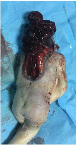
The first case is that of a 30-year-old female farmer with a notion of exposure to agricultural pesticides. She lives in a rural environment and has never attended school. She had no notable pathological antecedents, gravida 3 para 3 with the first two pregnancies not followed up leading to the vaginal birth of two healthy children at term. For the current pregnancy, she had only one antenatal visit at two months of pregnancy which showed an evolving intrauterine pregnancy. There was no folic acid supplementation in early pregnancy, nor was there any anaemia prophylaxis. She is blood group A Rhesus positive. Only HIV serology was negative. An obstetrical ultrasound was requested at the first consultation but not performed. She was referred to our clinic for premature rupture of membranes over 24 hours in a fetus presenting transversely in a pregnancy of unclear term. She complained of amniotic fluid loss associated with lumbopelvic pain. Clinical examination on entry noted good general condition, contractile uterus, uterine height 33 cm, empty pelvic excavation and absent fetal heart sounds. On vaginal touch the cervix is dilated to 3 cm, the membranes felt, the high presentation poorly appreciated. Obstetrical ultrasound on admission showed a progressive mono-fetal pregnancy of 31 weeks of amenorrhoea calculated by femoral measurement with a fetus in podalic presentation and positive fetal heart activity. Fetal head study revealed anencephaly associated with encephalocele and moderate hydramnios. The diagnosis was breech presentation in a fetus with anencephaly with encephalocele associated with moderate hydramnios at 31 weeks of amenorrhoea in the latent phase of labour. The vaginal delivery was directed for cervical dystocia of a polymalformed girl weighing 1400 g for a height of 33 cm. Macroscopic examination of the newborn showed an absence of the cranial vault, a batrachian facies (anencephaly), a membranous swelling at the upper end of the cephalic pole (encephalocele), an absence of neck development, and a total absence of skin covering along the spine (rachischisis). This complex malformation corresponds to a craniorachischisis (Figure 1). Macroscopic examination of the placenta revealed no abnormalities. The parents refused an autopsy.
The second case was that of a 24-year-old parturient, with no previous known medical condition, followed for her pregnancy in a public health center. This was her second pregnancy after a first spontaneous miscarriage. There was no folic acid supplementation in early pregnancy. Her blood type is O Rhesus positive. HIV, HBV, HCV, syphilis and toxoplasmosis serologies were negative. The obstetrical ultrasounds of the first and second trimester requested were not performed. She was referred to our facility for hydrocephalus in a pregnancy of 32 weeks of amenorrhea and 03 days with uterine contractions. The clinical examination on admission noted good general condition, flexible uterus, uterine height at 27 cm and fetal heart sounds present. On vaginal touch the cervix was mid-long open to 2 fingers with cephalic presentation. Obstetrical ultrasound on admission noted an evolving mono-fetal pregnancy with a cephalic fetus, positive cardiac activity and fetal weight estimate at 78th percentile. The cephalic pole was found to be significantly hydrocephalic with destruction of brain structures, the femoral length was small for the age of the pregnancy and the spine was not closed at the lower pole (spina bifida). The diagnosis was a threat of premature delivery with significant hydrocephalus associated with probable spina bifida in an evolving pregnancy at 32 weeks of amenorrhea and 03 days. The couple’s decision was to wait until birth. After the multidisciplinary meeting, their choice was granted and we did not start a tocolysis protocol but pulmonary maturation by corticotherapy was initiated. Vaginal birth was achieved during the night. The fetus was polymalformed male with macrocephaly, spina bifida, bilateral varus equinus club feet (Figures 2 and 3). The fetus weighed 1750 g with a head circumference of 44 cm and a height of 45 cm. Macroscopic examination of the placenta did not reveal any abnormalities. A karyotype test was requested from the couple but not carried out. The newborn died on the first day postpartum and the parents refused an autopsy.


Discussion
Neural tube defects (anencephaly and spina bifida) are very serious conditions, lethal in the case of anencephaly and disabling with psychomotor and neurological disorders in the case of spina bifida. The absence or insufficiency of folic acid (Vitamin B9) supplementation plays a major role in the occurrence of NTDs. Dietary insufficiency or deficiency and high multiparity are risk factors for folate deficiency and therefore factors for the occurrence of NTDs [4]. In our study, we found an absence of folic acid supplementation and a precarious situation. In addition, one of the patients presented an additional risk due to exposure to chemicals chemical products (agricultural fertilizers). This is a non-negligible exogenous factor likely to genetic alterations, particularly during gametogenesis. The role of folic acid in the prevention of NTDs is widely established in the literature. In Canada, the prevalence of NTDs in newborns has been declining since 1998 thanks to dietary diversification and the use of vitamin supplementation. However, there is still a risk of recurrence, which may indicate the involvement of genetic factors [4].
Thanks to technological advances, especially in the field of imaging, NTDs can be diagnosed in the first trimester of pregnancy by ultrasound. During prenatal follow-up, morphological ultrasound is recommended between 20 and 24 weeks of amenorrhea to detect fetal pathology [2]. Second trimester anatomical ultrasound with detailed assessment and imaging of the fetal intracranial and spinal areas is the primary screening test for fetal structural anomalies, including open/closed spinal dysraphism (anencephaly, encephalocele, spina bifida/myelomeningocele) [5]. The Society of Obstetricians and Gynaecologists of Canada detects more than 60% of morphological abnormalities on 2nd trimester ultrasound [6]. The RADIUS study shows an increase of more than 35% in the diagnosis of abnormalities before 24 weeks of amenorrhea in a level III unit and thus underlines the potential advantages of this type of structure in identifying major structural abnormalities in the fetus [6].
In our study, all cases were diagnosed late after 27 weeks of amenorrhea. This late ultrasound discovery could be explained by the lack of financial resources and awareness, which is responsible for late recourse to prenatal consultation and failure to carry out the requested assessment. Also, the cultural influence, which consists in delaying the declaration of pregnancy, has a negative impact on the early detection of malformations. It is essential to multiply communication sessions to change the population’s behavior. Hydramnios is a late ultrasound sign found in many cases of congenital malformations, particularly those of the nervous system.
Conclusion
Neural tube defects represent a pathology accessible to antenatal screening and whose management is well codified according to the term of the pregnancy. The delay in diagnosis of this condition is linked to the absence of early antenatal ultrasound due to the low socio-economic level of the couples concerned. The absence of folic acid supplementation is a risk factor. Thus, preventive measures are possible, including folic acid supplementation. Staff should work towards the establishment of a center for screening and treatment of fetal malformations. If detected early, the management of neural tube defects would be more adequate and limit late discovery in the third trimester.
Declarations
Guarantor of submission: corresponding author is the guarantor of submission.
Acknowledgements: None.
Funding: There are no funding sources to be declared.
Availability of data and materials: Supporting material is available if further analysis is needed.
Competing interests: The authors declare that they have no competing interests.
Consent for publication: Written informed consent was obtained from the patient for publication of this case report and any accompanying images. A copy of the written consent is available for review by the Editor-in-Chief of this journal.
Ethics approval and consent to participate: Ethics approval has been obtained to proceed with the current study. Written informed consent was obtained from the patient for participation in this publication.
References
- Charon P. Tératologie du tube neural: histoire et paléontologie. Antropo [Enligne]. 2005; 10.
- Heinrich W. Granier M. Les nouveaux marqueurs échographiques peuvent-ils améliorer le dépistage des défauts de fermetur
- Douglas R, Audibert F, Brock JA, et al. Anomalies foetales affectant le tube neural: Dépistage /diagnostic prénatal et prise en chargede la grossesse. J Obstet Gynaecol Can. 2014; 36: 940-942.
- Douglas W, Audibert F, Brock JA, et al. Supplémentation préconceptionnelle en acide folique/ mulivitamines pour la prévention primaire et secondaire des anomalies du tube neural et d’autres anomalies congénitales sensibles à l’acide folique. J Obstet Gynaecol Can. 2015; 37: S1- S19; 324.
- Douglas Wilson R. Anomalies foetales affectant le tube neural: Dépistage / diagnostic prénatal et prise en charge de la grossesse. Directive clinique de la sogc. 2016; 38: s496-s511.
- Gagnon A, Douglas W, Victoria M, et al. Evaluation des anomalies congénitales structurelles diagnostiquées pendant la période prénatale. JOGC. 2009; 31: 882–889.