
Journal of Clinical Images and Medical Case Reports
ISSN 2766-7820
Research Article - Open Access, Volume 3
Three-dimensional assessment of human enamel surfaces after adhesive resin removal using CLSM - an in vitro study
Babak Sayahpour1; Sara Eslami2*; Annika Ruhmann3; Sarah Buehling1; Abdolreza Jamilian4; Stefan Kopp1
1Department of Orthodontics, Johann-Wolfgang Goethe University, Frankfurt, Germany.
2Private Orthodontic Practice, Duesseldorf, Germany.
3Department of Conservative Dentistry, Johann-Wolfgang Goethe University, Frankfurt, Germany.
4Department of Orthodontics, Dental School, Cranio-Maxillofacial Research Center, Tehran Islamic Azad University of Medical Sciences, Iran.
*Corresponding Author : Sara Eslami
International Orthodontist, Grafenberger Allee 57, 40237, Duesseldorf, Germany.
Email: sara.eslami.sh@gmail.com
Received : May 18, 2022
Accepted : Aug 18, 2022
Published : Aug 25, 2022
Archived : www.jcimcr.org
Copyright : © Eslami S (2022).
Abstract
Objective: The aim of this study was to evaluate the effects of three different methods of adhesive resin removal on the enamel structure using a confocal laser scanning microscope (CLSM).
Methods: Initially, the enamel roughness of 39 extracted molars was determined using CLSM and the surface roughness parameters Sa, Sq and Sz were obtained. Subsequently, self-ligating metal brackets were applied using the composite Transbond XT. Group TC used a tungsten carbide bur to remove the adhesive residue, group TCP used a tungsten carbide bur to remove the majority of the adhesive remnant layer followed by an adhesive removal polisher to remove the rest of the layer and group TCF which used a tungsten carbide bur for the main adhesive removal and a fiberglass-reinforced bur for the final adhesive removal. Afterwards, the surface roughness was assessed again.
Results: There was a significant increase of the Sa and Sq parameters after using the tungsten carbide bur as the sole adhesive removal procedure. In groups TCP and TCF, the surface roughness values of Sa and Sq did not change significantly. In group TCF, the roughness even decreased with respect to the Sz parameter. The duration of adhesive removal was significantly greater in groups TCP and TCF than in group TC.
Conclusions: Our results suggest the superiority of the combination methods (TCP and TCF).
Keywords: Bracket; Adhesive; Bonding; Resin.
Citation: Sayahpour B, Eslami S, Ruhmann A, Buehling S, Jamilian A, et al. Three-dimensional assessment of human enamel surfaces after adhesive resin removal using CLSM - an in vitro study. J Clin Images Med Case Rep. 2022; 3(8): 2015.
Introduction
The introduction of the adhesive technique into orthodontics by Newmann in 1965 created the possibility of temporarily attaching brackets to the enamel [1]. An integral part of fixed orthodontic therapy is the appliance removal at the end of treatment which is achieved by inducing a bond failure using bracket removal pliers. An optimal bond failure occurs between the bracket base and the composite material, leaving adhesive residue on the teeth [2,3]. The remaining adhesive is not only esthetically unappealing, but it can also lead to discoloration and, more importantly, increase the risk of plaque accumulation. Therefore, this adhesive remnant should be removed.
Numerous studies have investigated different instruments for adhesive removal after bracket debonding [4-12]. These studies aimed to find an instrument that efficiently and gently polishes the enamel surface without leaving any roughness, such as ridges and grooves. Whilst instruments such as diamond burs 6-8, ultrasonic scalers [9,10] and lasers [6,11] have so far been proven inadequate or too destructive for adhesive removal, the use of tungsten carbide burs and subsequent polishing have shown promising results and are commonly recommended [10,12].
Their established place in everyday orthodontic practice has been shown in a survey by Webb et al. which demonstrated that the tungsten carbide bur is preferred by many orthodontists for composite removal [13]. This preference may be due to the efficiency of the tungsten carbide burs; these burs have sharp blades that enable them to remove adhesive remnants very quickly.
Other authors noted, however, an increase in surface roughness even with the use of the tungsten carbide burs [7,14]. Therefore, using these burs as the sole adhesive removal method could increase the surface roughness and, thus, could lead to a higher risk for plaque accumulation.
Another source of disagreement between the studies is the use of different methods for analyzing the surface roughness, such as contact-profilometers [14,15], atomic force microscopes (AFM) [16], 3D laser scanning devices [17] or scanning electron microscopes (SEM) [18,19]. Since the latter only produce high-resolution images of the enamel which require subjective evaluation, they cannot be recommended for making an observer’s independent statement. In contrast, the confocal laser scanning microscope (CLSM) can detect three-dimensional, extremely fine irregularities, making it capable of representing the degree of surface roughness quantitatively by a numerical value.
The aim of the present in vitro study was to determine the effects of three adhesive removal methods on the enamel surface roughness by using CLSM. The methods included an abrasive set (Smoozie Set, Komet Dental) and a fiberglass-reinforced bur (Stainbuster®, Abrasive Technology).
Materials and methods
This in vitro study was performed on 39 human maxillary and mandibular molars that had been previously extracted for other medical reasons.
The teeth were stored immediately after extraction in dis tilled water at room temperature. Only teeth with an intact buccal surface were included in the study. Molars that showed damage to the tooth structure, enamel cracks, demineralization or restorations were excluded from the study.
The buccal enamel surfaces of the teeth were cleaned for 10 seconds each, using polishing cups (Prophy Brushes, Henry Schein, Melville, New York, US) and fluoride-free polishing paste (Zircate Prophy Paste, Dentsply DeTrey GmbH, Germany). Afterwards, the residue of the paste was thoroughly removed under water spray.
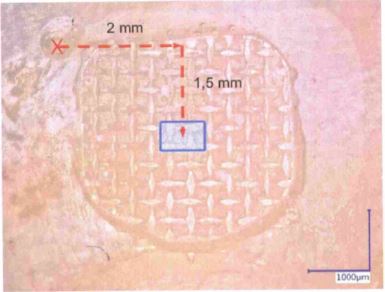
The teeth were then embedded in resin blocks, leaving the buccal tooth surfaces free of embedding material. A reference hole was drilled into each buccal surface to ensure the reproducibility of the sample positioning under the microscope (Figure 1).
The samples were then positioned under the microscope. Subsequently, the enamel surface roughness of the specimens was quantitatively determined using the Keyence VK-X100 confocal laser scanning microscope (Keyence Corporation, Osaka, Japan) and the surface roughness parameters Sa (arithmetical mean height), Sq (root mean square height) and Sz (maximum height) were recorded (T0 measurements). For this purpose, the analysis program VK-Analyzer was used. The investigated enamel surfaces had dimensions of 500 x 700 μm and were measured by a 20 x optical system.
The buccal enamel surfaces were then conditioned with phosphoric acid (Ultra Etch, Ultradent Products Inc., US) for 20 seconds. Subsequently, the acid was thoroughly removed using water spray and the enamel surface dried via an oil-free air spray. Transbond™ XT Primer (3M Unitek, California, US) was applied thinly with a microbrush to the enamel surface. The Transbond™ XT composite (3M Unitek, California, US) was then applied to the bracket base (Damon Q, Ormco, California, US) in an even layer thickness and the bracket was placed beneath the reference hole on the buccal tooth surface under light pressure. The excess adhesive was carefully removed. Subsequently, the composite was cured using a polymerization lamp (Bluephase Style, Ivovlar Vivadent, Schaan, Liechtenstein) on the mesial and distal sides for 10 seconds each at a distance of 5 mm from the enamel surface.
After bracket application, we stored the specimens in a water bath at a temperature of 37°C for 24 hours. The brackets were then debonded by a single, experienced orthodontist using bracket removal pliers (Damon Q Debonding Instrument, Ormco, California, US).
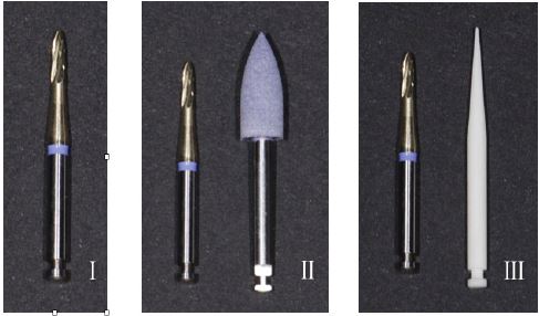
Subsequently, the specimens were randomly assigned to three groups of 13 teeth each. In group TC, the tungsten carbide bur of the Smoozie set (Komet Dental, Lemgo, Germany) was used as a single-step method to remove the adhesive remnants. In group TCP, a two-step procedure was employed in which the majority of the adhesive remnant layer was reduced using the tungsten carbide bur. As soon as only a thin adhesive layer remained and there was a risk of abrading the underlying enamel, the adhesive removal polisher of the Smoozie Set (Komet Dental, Lemgo, Germany) was used. A similar approach was used in group TCF. Instead of adhesive removal polisher, a fiberglass-reinforced bur (Stainbuster, Abrasive Technology, Lewis Center, Ohio, US) was used to remove the final layer of adhesive remnants (Figure 2). The instruments were used with water cooling and the composite removal was performed by one, single researcher.
The roughness parameters Sa, Sq and Sz were recorded again following the adhesive removal (T1 measurements). The time required to remove the adhesive residue was recorded for ten test specimens from each group.
Statistical analysis was performed using the BiAS program (Epsilon Verlag, Germany). The significance level was set at p ≤ 0.05. The Shapiro-Wilk test showed that no normal distribution of the data could be assumed; for this reason, non-parametric test procedures, such as the Wilcoxon matched-pairs test and the Kruskal-Wallis test, were applied. For post-hoc analysis, multiple Conover-Iman tests were performed. A Bonferroni-Holm correction was used to reduce the statistical probability of error.
Results
The results of the roughness measurements are summarized in Tables 1-3. Group TC showed a significant increase in surface roughness with respect to the parameters Sa and Sq. On the contrary, no significant differences were observed in the roughness of the enamel surfaces in groups TCP and TCF. The surface roughness was even reduced in group TCF with respect to the Sz parameter. The results of the Kruskal-Wallis test, shown in Table 4, indicated that group TC differed significantly from groups TCP and TCF (p ≤ 0.05) with regard to the surface roughness after adhesive removal, whereas no significant differences could be found when comparing groups TCP and TCF.
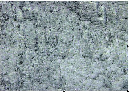
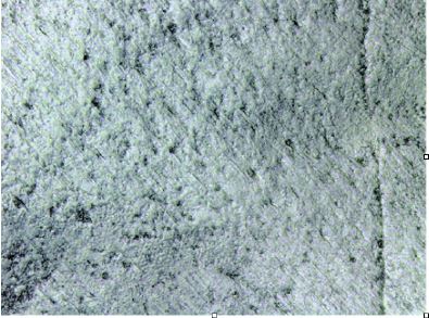
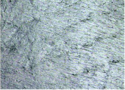
Table 1: Roughness pattern after adhesive removal using the tungsten carbide bur followed by the fiberglass-reinforced bur (group TCF).
| Group TC | ||||
|---|---|---|---|---|
| Parameter | Measurement | Median | Q1 - Q3 | p-value |
| Sa | T0 | 0.572 | 0.533 - 0.643 | 0.0002* |
| T1 | 0.805 | 0.759 - 0.856 | ||
| Sq | T0 | 0.757 | 0.705 - 0.862 | 0.0002* |
| T1 | 1.073 | 1.003 - 1.179 | ||
| Sz | T0 | 14.087 | 10.506 - 18.057 | 0.06 |
| T1 | 19.265 | 15.956 - 23.049 | ||
Table 2: Surface roughness parameters in group TCP before (T0) and after (T1) adhesive removal in μm.
| Group TCP | ||||
|---|---|---|---|---|
| Parameter | Measurement | Median | Q1 - Q3 | p-value |
| Sa | T0 | 0.601 | 0.549 - 0.715 | 0.95 |
| T1 | 0.590 | 0.535 - 0.666 | ||
| Sq | T0 | 0.840 | 0.756 - 0.961 | 1.00 |
| T1 | 0.780 | 0.709 - 0.888 | ||
| Sz | T0 | 17.276 | 14.952 - 18.960 | 0.15 |
| T1 | 12.768 | 10.874 - 17.604 | ||
Table 3: Surface roughness parameters in group TCF before (T0) and after (T1) adhesive removal in μm.
| Group TCF | ||||
|---|---|---|---|---|
| Parameter | Measurement | Median | Q1 - Q3 | p-value |
| Sa | T0 | 0.633 | 0.558 - 0.747 | 0.38 |
| T1 | 0.600 | 0.538 - 0.672 | ||
| Sq | T0 | 0.832 | 0.742 - 1.130 | 0.17 |
| T1 | 0.787 | 0.707 - 0.881 | ||
| Sz | T0 | 17.752 | 14.509 - 29.748 | 0.0012* |
| T1 | 14.210 | 11.303 - 15.430 | ||
Table 4: Results of the Kruskal-Wallis test followed by multiple Conover-Iman tests comparing the surface roughness parameters between each treatment group.
| Group | Comparison group | p-value | |
|---|---|---|---|
| DSa | TC TC TCP |
TCP TCF TCF |
0.0000* 0.0000* 0.58 |
| DSq | TC TC TCP |
TCP TCF TCF |
0.0000* 0.0000* 0.32 |
| DSz | TC TC TCP |
TCP TCF TCF |
0.0096* 0.0004 0.21 |
Table 5: The required time for adhesive removal (in seconds) for each treatment group.
| Group | n | Mean value | Minimum | Maximum |
|---|---|---|---|---|
| TC | 10 | 58.7 | 48.5 | 65.9 |
| TCP | 10 | 84.1 | 66.9 | 114.2 |
| TCF | 10 | 74.4 | 56.2 | 96.4 |
Table 6: Comparison of the groups with regard to the required time for adhesive removal.
| n | Minimum | Maximum | |
|---|---|---|---|
| Time | TC | TCP | 0.0001* |
| TCP | TCF | 0.0035* | |
| TCF | TCF | 0.14 |
Visual differences between the groups could also be observed in the images obtained with the confocal laser scanning microscope. While the tungsten carbide bur had left a coarse roughness pattern (Figure 3), the adhesive removal polisher and fiberglass-reinforced bur were able to produce more homogeneous and smoother enamel surfaces (Figures 4 and 5).
Table 5 shows the average time required for adhesive removal. Group TC was, as predicted, the fastest method, requiring only 59 seconds on average. The groups TCF and TCP required significantly more time, with an average of 74 and 84 seconds, respectively. According to the Kruskal-Wallis test, no significant differences were found between the groups TCP and TCF regarding the required time for adhesive removal (Table 6).
Discussion
The present study investigated the effect of three adhesive removal procedures on the enamel surface roughness by using CLSM.
A major advantage of the present study is the use of CLSM as a non-destructive objective measurement tool to perform before and after measurements on the same sample. CLSM requires minimal sample preparation and can obtain both two- and three-dimensional images simultaneously. Contrary to SEM, which has been used in many other studies, CLSM is able to perform quantitative objective roughness analysis without requiring the subjective assessment of enamel quality.
Another pitfall of some studies is the incorporation of indices, such as the Enamel Surface Index (ESI) or the Enamel Damage Index (EDI), for surface evaluation after bracket debonding [20-22]. These indices need a subjective assessment by an examiner and, therefore, cannot be recommended for an objective evaluation.
Various methods of adhesive removal after bracket debonding are described in the literature [23-25]. Among these methods, tungsten carbide burs have established their place as the method of choice in everyday orthodontic practice [13,26]. However, this method leads to irreversible changes in the enamel structure [7,14]. This drawback was also confirmed in our study using the tungsten carbide bur of the Smoozie Set as the sole adhesive removal method in group TC. As can be seen in Figure 3, the bur left an uneven and inhomogeneous enamel surface. According to the statistical evaluations, there was a significant increase of the roughness parameters Sa and Sq.
A study by Karan et al. [16] compared a tungsten carbide bur with the application of a fiberglass-reinforced bur. In contrast to our study, the fiberglass-reinforced bur was used as the sole method of adhesive removal and was not combined with the tungsten carbide bur. Although homogeneous enamel surfaces were achieved with this method, the required time for adhesive removal was twice as long as with the use of the tungsten carbide bur. A combined two-step method (as used in the present study in groups TCP and TCF) creates a homogeneous enamel surface without being too time consuming.
Only a few studies have investigated a two-step combined procedure in which, first, a coarse and then a fine bur is applied. Our findings support the use of combined methods, such as TCP or TCF, since the enamel surfaces were not significantly influenced in these groups regarding the parameters Sa and Sq; method TCF has even led to a reduction of surface roughness with respect to the Sz parameter.
Method TC was significantly faster than the other groups since only one stage of adhesive removal was performed in this group. Other studies have also found the tungsten carbide burs to be convincing due to their rapid operation [11]. In the study of Degrazia et al., a 30-bladed tungsten carbide bur was used [27]; here, the duration of composite removal was similar to our study (59.2 seconds), whereas when a 5-bladed tungsten carbide bur was used, 75.5 seconds were needed. Furthermore, compared to method TC, methods TCP and TCF require more time (84 and 74 seconds, respectively). The difference of 15-25 seconds is statistically significant, but is still clinically acceptable.
Conclusion
Finally, it can be concluded that the use of the tungsten carbide bur in combination with the adhesive removal polisher or the fiberglass-reinforced bur can be recommended as both of these methods did not significantly increase the enamel roughness and are not too time consuming. The tungsten carbide bur, although convincing in its rapid operation, resulted in an increase in surface roughness and, thus, cannot be recommended.
In practice, the attempt to remove the adhesive remnants in a single-step method using tungsten carbide burs can create a rough appearance which then necessitates a subsequent polishing procedure with pumice powder or a polishing paste. Since this additional working step is almost always necessary, the use of these burs shows no advantage over the two-step procedures of groups TCP and TCF.
Further studies are required which, in addition to the homogeneity of the tooth surfaces, should also investigate the enamel loss caused by the removal of the adhesive residue.
Declarations
Ethics approval and consent to participate: Ethical approval for this study was obtained from the ethics committee at the Johann-Wolfgang Goethe University (approval number: 19-264).
Consent for publication: I, Sara Eslami and the other authors, give our consent for the publication of identifiable details, which can include photographs and details within the text (“Material”) to be published.
Availability of supporting data: The data that support the findings of this study are available on request from the corresponding author.
Competing interests: The Authors declare, that they have no financial or non-financial competing interests.
Funding: The study was funded by the orthodontic department of the Johan-Wolfgang Goethe University.
Acknowledgements: Not applicable
References
- Newman GV (1965). Epoxy adhesives for orthodontic attachments: progress report. Am J Orthod. 51: 901-912.
- Fan XC, Chen L, Huang XF (2017). Effects of various debonding and adhesive clearance methods on enamel surface: an in vitro study. BMC Oral Health. 17:58.
- Pont HB, Ozcan M, Bagis B, Ren Y (2010). Loss of surface enamel after bracket debonding: an in-vivo and ex-vivo evaluation. Am J Orthod Dentofacial Orthop. 138: 387. e381-387.e389.
- Janiszewska-Olszowska J, Szatkiewicz T, Tomkowski R, Tandecka K, Grocholewicz K (2014). Effect of orthodontic debonding and adhesive removal on the enamel - current knowledge and future perspectives - a systematic review. Med Sci Monit. 20: 1991-2001.
- Tonetto M, Frizzera F, Porto T, Jordão K, dos Santos R, de Andrade M, Klug R, Bandιca M (2014). Methods for removal of resin remaining after debonding of orthodontic brackets: a literature review. J Dent Res Rev. 1: 105-107.
- Ahrari F, Akbari M, Akbari J, Dabiri G (2013). Enamel surface roughness after debonding of orthodontic brackets and various clean-up techniques. J Dent (Tehran). 10: 82-93.
- Eliades T, Gioka C, Eliades G, Makou M (2004). Enamel surface roughness following debonding using two resin grinding methods. Eur J Orthod. 26: 333-338.
- Hong YH, Lew KK (1995). Quantitative and qualitative assessment of enamel surface following five composite removal methods after bracket debonding. Eur J Orthod. 17: 121-128.
- Hosein I, Sherriff M, Ireland AJ (2004). Enamel loss during bonding, debonding, and cleanup with use of a self-etching primer. Am J Orthod Dentofacial Orthop. 126: 717-724.
- Ireland AJ, Hosein I, Sherriff M (2005). Enamel loss at bond-up, debond and clean-up following the use of a conventional light-cured composite and a resin-modified glass polyalkenoate cement. Eur J Orthod. 27:413-419.
- Yassaei S, Aghili H, Joshan N (2015). Effects of removing adhesive from tooth surfaces by Er:YAG laser and a composite bur on enamel surface roughnessand pulp chamber temperature. Dent Res J (Isfahan). 12:254-259.
- Zachrisson BU, Arthun J (1979). Enamel surface appearance after various debonding techniques. Am J Orthod. 75: 121-127.
- Webb BJ, Koch J, Hagan JL, Ballard RW, Armbruster PC (2016) Enamel surface roughness of preferred debonding and polishing protocols. J Orthod. 43: 39-46.
- Garg R, Dixit P, Khosla T, Gupta P, Kalra H, Kumar P (2018). Enamel surface roughness after debonding: a comparative study using three different burs. J Contemp Dent Pract. 19: 521-526.
- erreira FG, Nouer DF, Silva NP, Garbui IU, Correr-Sobrinho L, Nouer PR (2014). Qualitative and quantitative evaluation of human dental enamel after bracket debonding: a noncontact three-dimensional optical profilometry analysis. Clin Oral Investig. 18: 1853-1864.
- Karan S, Kircelli BH, Tasdelen B (2010). Enamel surface roughness after debonding. Angle Orthod. 80: 1081-1088.
- Brauchli LM, Baumgartner EM, Ball J, Wichelhaus A (2011). Roughness of enamel surfaces after different bonding and debonding procedures: an in vitro study. J Orofac Orthop. 72: 61-67.
- Gracco A, Lattuca M, Marchionni S, Siciliani G, Alessandri Bonetti G (2015). SEM-Evaluation of enamel surfaces after orthodontic debonding: a 6 and 12-month follow-up in vivo study. Scanning 37: 322-326.
- Radlanski RJ (2001). A new carbide finishing bur for bracket debonding. J Orofac Orthop. 62:296-304.
- Alessandri Bonetti G, Zanarini M, Incerti Parenti S, Lattuca M, Marchionni S, Gatto MR (2011). Evaluation of enamel surfaces after bracket debonding: an in-vivo study with scanning electron microscopy. Am J Orthod Dentofacial Orthop. 140: 696-702.
- Baumann DF, Brauchli L, van Waes H (2011). The influence of dental loupes on the quality of adhesive removal in orthodontic debonding. J Orofac Orthop. 72: 125-132.
- Sessa T, Civovic J, Pajevic T, Juloski J, Beloica M, Pavlovic V, Glisic B (2012). Scanning electron microscopic examination of enamel surface after fixed orthodontic treatment: in-vivo study. Srp Arh Celok Lek. 140: 22-28.
- Amasyalı M, Sabuncuoğlu FA, Ersahan Ş, Oktay EA (2019). Comparison of the effects of various methods used to remove adhesive from tooth surfaces on surface roughness and temperature changes in the pulp chamber. Turk J Orthod. 32: 132-138.
- Atabek D, Sungurtekin Ekçi E, Bani M, Öztaş N (2016). The effect of various polishing systems on the surface roughness of composite resins. Acta Odontol Turc. 33: 69-74.
- Cardoso LA, Valdrighi HC, Vedovello Filho M, Correr AB (2014). Effect of adhesive remnant removal on enamel topography after bracket debonding. Dental Press J Orthod. 19: 105-112.
- Campbell PM (1995). Enamel surfaces after orthodontic bracket debonding. Angle Orthod. 65: 103-110.
- Degrazia FW, Genari B, Ferrazzo VA, Santos-Pinto AD, Grehs RA. Enamel Roughness Changes after Removal of Orthodontic Adhesive. Dent J (Basel). 2018; 6(3): 39.