
Journal of Clinical Images and Medical Case Reports
ISSN 2766-7820
Clinical Image - Open Access, Volume 3
Dental calculus contributing to impaired
speech and deglutition: A rare case
*Corresponding Author : Aparna Kaushik
Department of Periodontology, Post Graduate Institute of Dental Sciences, Rohtak, Haryana, India.
Email: aparna81112@gmail.com
Received : Aug 08, 2022
Accepted : Aug 30, 2022
Published : Sep 06, 2022
Archived : www.jcimcr.org
Copyright : © Aparna K (2022).
Abbreviations: GI: Gingival Index; PPD: Probing Pocket Depth; CAL: Clinical Attachment Level.
Citation: Kaushik A. Dental calculus contributing to impaired speech and deglutition: A rare case. J Clin Images Med Case Rep. 2022; 3(9): 2033.
Case description
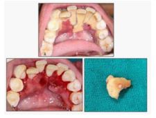
A 21 year female patient reported with the chief complaint of pain and difficulty while eating and swallowing food from past 15-20 days. Her mother complained about her silent mode most of the time. Patient revealed that she is having pain while speaking, therefore she avoid speaking from past few days. Patient was systemically healthy. Intraoral examination revealed plaque, heavy calculus deposits and bleeding on probing. In mandibular anterior teeth, calculus deposits extended till the lingual vestibule with respect to right mandibular central incisor (Figure 1a). Severe gingival inflammation was there. Calculus was irritating the lingual floor mucosa leading to pain while swallowing and speaking. Calculus contributes indirectly to gingival inflammation by providing fixed nidus to the microbial plaque, primary etiologic agent, for its retention near the periodontal tissues [1]. Mean GI [2], PPD and CAL were 3, 1.9 and 2.5 respectively for mandibular anteriors.
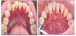
Patient was made comfortable at the dental chair. Phase I therapy aiming removal of plaque biofilm, calculus was performed using ultrasonic scaler. After scaling, a soft tissue deformity on the lingual side of mandibular anteriors was visible (Figure 1b). Calculus fragment is here depicted in figure 1c. Patient was educated about her condition. Brushing technique [3] was demonstrated. Twice daily brushing using ultrasoft toothbrush was recommended. Topical benzocain gel and 0.2% clorhexidine mouthwash twice daily were prescribed for two weeks. After one week, patient reported 80% relief in pain and discomfort (Figure 2a). She started talking normally again. Full mouth scaling was done. Patient was motivated and oral hygiene was reinforced. Healing of soft tissue defect was noted at one month (Figure 2b). Patient was satisfied and didn’t reported for further treatment.
References
- Carranza F, Newman M, Takei H, Klokkevold P. Carranza’s Clinical Periodontology. 11th ed. St. Louis, Mo.: Elsevier Saunders. 2012;
- Loe H, Silness J. Periodontal Disease I pregnancy. I. Prevalence And Severity. Acta odontologica Scandinavica. 1963; 21: 533–551. https://doi.org/10.3109/00016356309011240
- Bass CC. An effective method of personal oral hygiene. Part II. J la State Med Soc. 1954; 106: 100.