
Journal of Clinical Images and Medical Case Reports
ISSN 2766-7820
Clinical Image - Open Access, Volume 3
Asymptomatic disseminated cysticercosis:
A rare incidental finding
Prabhat*; Ananya; Mayank; Rohit; Ravi Kant; Monika
Department of Internal Medicine, All India Institute of Medical Sciences, Rishikesh,Uttarakhand, India.
*Corresponding Author : Prabhat Rijal
Department of Internal Medicine, All India Institute of Medical Sciences, Rishikesh,Uttarakhand, India.
Email: prijal103@gmail.com
Received : Aug 03, 2022
Accepted : Aug 30, 2022
Published : Sep 06, 2022
Archived : www.jcimcr.org
Copyright : © Rijal P (2022).
Citation: Rijal P, Ananya, Mayank, Rohit, Kant R, et al. Asymptomatic disseminated cysticercosis: A rare incidental finding. J Clin Images Med Case Rep. 2022; 3(9): 2034.
Background
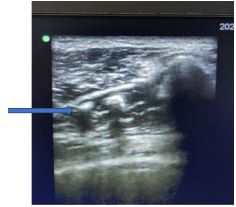
A middle-aged male, coming from a remote hilly village of Northern India, presented to the outpatient department for optimization of his anti-diabetic medications. On routine general examination innumerable soft to firm, asymptomatic subcutaneous nodular swellings were noted over his trunk and limbs which varied from 1-4 cm in size. However, he denied any history of headache, seizures, body ache, muscle pains, fever, or systemic involvement. He stated that he occasionally consumed pork since childhood.
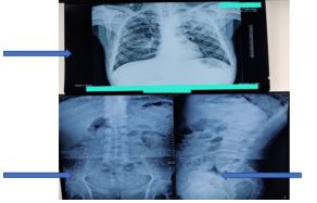
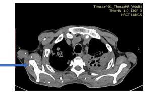
Baseline investigations including complete blood count, liver and renal function tests, stool analysis and electrocardiogram were within normal limits. On further evaluation chest and pelvic roentgenogram showed multiple homogenous small sized opacites in the muscular planes. A contrast enhanced MRI Brain and Spine showed multiple paraspinal and cortical cysticercosis with calcification and no active lesions. Fundoscopy did not reveal ocular cysticercosis. A diagnosis of asymptomatic disseminated Myocysticercosis with neurocysticercosis was made. Since the cysticerci were already calcified, no active treatment was given.
Human cysticercosis caused by larva of Taenia solium, is a major public health problem in developing countries with Neurocysticercosis being the most common neuroparasitic infection [1]. Myocysticercosis is second most common site of cysticercosis. Magnetic resonance imaging remains the best modality for diagnoses and can identify various stages of the parasite. Disseminated Cysticercosis is a rare occurrence, that is caused by Hepato-portal dissemination of the larva of T. Solium to various organs [2]. Patients usually present with complains of headache, seizures or systemic complains [3]. However asymptomatic disseminated cysticercosis has been reported in only a handful of cases. Hence, we present a case of incidentally diagnosed disseminated cysticercosis with classical imaging findings.
References
- Prasad KN, Prasad A, Verma A, Singh AK. Human cysticercosis and Indian scenario: A review. Journal of Biosciences. 2008; 33: 571-582.
- Jawale R, Duberkar D. Disseminated cysticercosis. Neurology. 2015; 84: 327.
- Khandpur S, Kothiwala SK, Basnet B, Nangia R, Venkatesh HA, Sharma R. Extensive disseminated cysticercosis. Indian J Dermatol Venereol Leprol. 2014; 80: 137-140.