
Journal of Clinical Images and Medical Case Reports
ISSN 2766-7820
Short Report - Open Access, Volume 3
When the dust has settled…An occupational disease with swelling of the cervical lymph nodes
Philipp Kulas1*; Wolfgang Tränkenschuh2; Alessandro Bozzato1; Dragan Kopanja1; Lukas Pillong1
1Department of Otorhinolaryngology, Head and Neck Surgery, University Hospital Saarland Medical School, Hom-burg, Germany
2Department of Pathology, University Hospital Saarland Medical School, Homburg, Germany.
*Corresponding Author : Kulas Philipp
Department of Otorhinolaryngology, Saarland University Medical Center, Kirrberger Strasse, 66421, Homburg/Saar, Germany.
Email: philipp.kulas@uks.eu
Received : Aug 11, 2022
Accepted : Sep 02, 2022
Published : Sep 09, 2022
Archived : www.jcimcr.org
Copyright : ©Philipp K (2022).
Keywords: Lymphadenopathy; Silicosis; Sarcoidosis; Occupational disease; Dental technician.
Citation:Kulas P, Tränkenschuh W, Bozzato A, Kopanja D, Pillong L. When the dust has settled…An occupational disease with swelling of the cervical lymph nodes. J Clin Images Med Case Rep. 2022; 3(9): 2043.
Introduction
Cervical lymphadenopathy is a common finding in the everyday life of an ORL physician. The diagnostic steps range from laboratory and microbiological examinations to removing lymph nodes for histopathological processing. The differential diagnoses include infectious, chronic and malignant diseases. In rare cases, lymph node biopsy may diag-nose a chronic lung disease like sarcoidosis or silicosis.
In the following, we report on the case of a dental technician with the suspected diagnosis of sarcoidosis. Because of therapy-refractory cervical lymphadenopathy, a node removal was performed leading to the diagnosis of silicosis and subsequently to recognition as an occupational disease.
Case report
A 43-year-old male patient presented to our tertiary care center with a painless right supraclavicular neck swelling that had been present for more than 12 months. The patient also described progressive exertional dyspnea in connection with a feeling of tightness in the thoracic region that had existed for about four months and a non-productive cough. B symptoms like fever, weight loss, night sweats or exhaustion were denied.
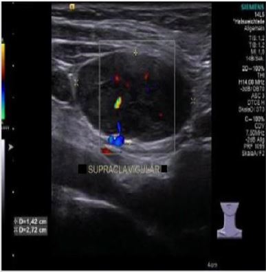
Nicotine use had been discontinued three years ago (cumulative 20 pack-years). The patient worked as a dental technician and otoplastist (total 25 years). Six years ago, a bronchoalveolar lavage had been performed with a suspected diagnosis of sarcoidosis due to similar symptoms and bihilary lymphadenopathy. However, this hypothesis could not be confirmed. A mobile, indurated lymph node could be palpated on the right supraclavicular neck region at our clinical examination. The duplex sonography exam revealed a round, sharply demarcated, homogeneous, anechoic mass measuring 3.76 x 1.42 x 2.72 cm with a central vascularisation pattern most likely to be an enlarged lymph node (Figure 1).
The other ORL findings were normal. The CT scan images showed a progression of the bihilary lymphadenopathy compared to the previous examinations six years ago. Furthermore, a new, low-grade involvement of the lungs by predominantly submiliary foci in a perilymphatic distribution was shown. Corticosteroid therapy was initiated but did not improve the symptoms. As a final consequence, the patient was presented to our department because the suspected diagnosis of sarcoidosis had not been confirmed, but symptoms progressed. A lymph node excision under general anesthesia was completed. There was no evidence of sarcoidosis or malignant disease in the histopathological work-up. The tissue showed increased iron storage, nodular fibrosis zones with embedded foreign material and overall lympho-follicular hyperplasia (Figure 2). In conjunction with the professional history of a dental technician, the finding was suspicious of dust-associated lymphadenopathy or lymph node silicosis. As a result, an application was made to recognize an occupational disease, which has now also been confirmed. The patient has left her original job and has retrained.

Discussion
Interstitial lung diseases caused by inhaling mixed inorganic dust are generally referred to as pneumoconioses [1]. A distinction is made between quartz dust silicosis caused by inhalation of almost pure quartz dust and mixed dust silicosis being driven by additional inhalation of dust mixtures of different compositions. However, the term “silicosis” in the narrower sense only describes pneumoconiosis that is caused by crystalline types of dust with a high concentration of silica.
Silicosis has been listed and recognized as an occupational disease in Germany under the number BK 4101 since 1929. While initially the “dust lung” was primarily associated with occupational groups from hard coal and ore mining, today there is also clear evidence of illnesses during employment, as in the case described, in dental laboratories [2].
For 2018, 1116 notifications and 495 recognitions of a BK no. 4101 were documented (DGUV).
The factors that influence the development of silicosis include the dust concentration in the breath, the duration of exposure, the proportion of the respirable dust fraction, the individual’s ability to clean the lungs and other specific properties of the dust. The silicosis is often symptom-free in the early stages or is occasionally noticeable as exertional dyspnea. Over time, however, a progressive course with the development of respiratory insufficiency, pulmonary hypertension, and possibly right heart failure in the sense of a cor pulmonale is possible [3].
It should also be noted that there is an increased risk of bronchial carcinoma, pulmonary tuberculosis or other myco-bacterial diseases, and spontaneous pneumothorax.
In the differential diagnosis, as in the case described above, sarcoid and other granulomatous lung changes must be considered.
An extrathoracic or cervical manifestation, as in this case, was previously described once by Tze-Wai Wong and colleagues in 1998 [4]. It is reported by a 54-year-old employee of a coal plant who, as a secondary finding when treating a femoral fracture, had supraclavicular lymphadenopathy. After securing bioptic material, the diagnosis was made based onthe occupational history. Imaging procedures performed years earlier for unexplained dyspnea could help confirm the diagnosis.
Conclusion
In unclear, persistent lymphadenopathy cases, a histopathological examination in the sense of lymph node extirpation is frequently required to exclude malignancy.
A detailed anamnesis with clarification of the environmental surroundings in the case of unspecific complaints and symptoms in a combination of lymph node enlargement is essential.
Finally, the interdisciplinary communication between the institutions, especially with the Institute for Pathology, as well as radiology and pulmonology departments, may lead to the final diagnosis of silicosis. Merely naming the tissue sent in for histopathological examination is rarely helpful. In the case described above, the suspected diagnosis of an occupational disease could only be made by specifying the occupational history and the individual clinical course, thus eliminating the most varied differential diagnoses that had existed for years.
References
- Ulmer WT, Reichel G. Pneumokoniosen. 1976.
- Wang X, Yu ITS, Wong TW, Yano E. Respiratory symptoms and pulmonary function in coal miners: Looking into the effects of simple pneumoconiosis. Am J IndMed. 1999; 35: 124–131.
- Baur X, Heger M, Bohle R, Hering K, Hofmann-Preiß K, Nowak D, et al. S2k-Leitlinie nach AWMF-Schema der Deutschen Gesellschaft für Pneumologie und Beatmungsmedizin und der Deutschen Gesellschaft für Arbeitsmedizin und Umweltmedizin “Diagnostik und Begutachtung der Berufskrankheit Nr. 4101 Quarzstaublungenerkrankung (Silikose) 1 der Berufskrankheitenverordnung.” Pneumologie. 2016; 70: 782–812.
- Wai WONG T, Sun YU T, Ming TAM C, Rong WANG X. A silicosis patient presenting with an enlarged supra-clavicular lymph node. Respirology. 1998; 3: 281–283.