
Journal of Clinical Images and Medical Case Reports
ISSN 2766-7820
Case Report - Open Access, Volume 3
Postpartum vulvar expansive hematoma:
An uncommon case report
Aziz Slaoui*; Mariam Mahtate; Madiha Ouchamch; Najia Zeraidi; Amina Lakhdar; Aziz Baydada; Aicha Kharbach
Gynaecology-Obstetrics and Endoscopy Department, Maternity Souissi, University Hospital Center IBN SINA, University Mohammed V, Rabat, Morocco.
*Corresponding Author : Aziz Slaoui
Gynaecology-Obstetrics and Endoscopy Department & Gynaecology-Obstetrics and Endocrinology Department, University Mohammed V, Rabat, Morocco.
Email: azizslaoui27@gmail.com
Received : Aug 11, 2022
Accepted : Sep 05, 2022
Published : Sep 12, 2022
Archived : www.jcimcr.org
Copyright : © Slaoui A (2022).
Abstract
Background: Postpartum hemorrhage is the leading world cause of maternal mortality in low-and-middle income countries. Puerperal hematomas are auncommon cause of postpartum hemorrhage. Their adequate management requires a particular skill and technical platform to surgically assure hemostasis. This case allows us to draw the attention of practitioners to the seriousness and singularity of this highly morbid pathology.
Case presentation: We hereby report the case of a 22-year-old woman, G2P2 with a live child, hospitalized in our structure for the management of a vaginal delivery that was complicated by a hemorrhagic shock that occurred one hour later. She was then treated surgically for an expansive vulvar hematoma.
Conclusions: This case allows us to draw the attention of practitioners to the seriousness and singularity of puerperal hematomas. It is an uncommon but serious postpartum complication. It can be associated with a state of shock which can threaten the vital prognosis of the parturient. Practitioners should be aware that the outcome depends on the promptness of diagnosis and management.
Keywords: Postpartum hemorrhage; Puerperal hematomas; Vaginal delivery complications.
Citation: Slaoui A, Mahtate M, Ouchamch M, Zeraidi N, Lakhdar A, et al. Postpartum vulvar expansive hematoma: An uncommon case report. J Clin Images Med Case Rep. 2022; 3(9): 2045.
Background
Postpartum hemorrhage is the leading world cause of maternal mortality with a contribution ranging from 18 to 50% of deaths in low-and-middle income countries [1-5]. The most common etiologies are: uterine atony, coagulopathies, retained placental debris, placental insertion anomalies, retained fetal membranes and genital tract lacerations [4,5]. Unusual causes include puerperal hematomas (also called peri-genital thrombi) with a frequency of 1/1000 deliveries [6]. The latter are easily diagnosed when they involve the vulva and/or vagina, whereas pelvic (retroperitoneal) locations require more subtlety on the part of the practitioner as well as the performance of imaging examinations [7]. We hereby report the uncommon case of an expansive vulvar hematoma in the immediate postpartum period, managed surgically.
Conclusion
We report the case of a young woman aged 22 years, gravida 2 para 2, who was admitted in our department for vaginal delivery. She had no particular medical antecedents and her first pregnancy had resulted in a full-term vaginal delivery without complications two years earlier, of a daughter weighing 3200 grams. She has never received a blood transfusion and was blood group B, rhesus positive.
After her instrumental delivery assisted by vacuum cup with active placental delivery without any particularity, we noted a vaginal bleeding of great abundance estimated at 800 cc. Postpartum hemorrhage was diagnosed and we immediately initiated: uterine massage, uterine revision (which was negative), infusion of 5% glucose serum containing 40 international units of oxytocin with then 1g of tranexamic acid. When the vaginal bleeding stopped, extreme asthenia and mucocutaneous pallor were observed, consistent with an arterial hypotension at 71/43 millimeters of mercury, as well as the appearance a few minutes later of progressive and very painful swelling of the left labia majora.
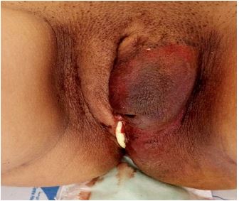
On physical examination, the abdomen was soft and painless. Hydro-aeric noises were present and the uterus was well contracted. Inspection of the external genitalia revealed a bluish swelling of the left hemivulva reaching the inguinal crease externally, the gluteal maxima posteriorly, the venus mount anteriorly. Internally, the mass pushed back the labia minora and protruded into the vaginal wall from which a fine trickle of blood flowed through a tear involving the muscularis and the mucosa (Figures 1 and 2). The skin was shiny, under great tension, and had patches of dermabrasion. Palpation was extremely painful and made it difficult to explore the mass without anesthesia. Its large diameter was anteroposterior and measured 16 centimeters while its small diameter, transverse, measured 13 centimeters. These dimensions increased quite rapidly as did the pain reported by the patient. We made the diagnosis of an immediate postpartum expansive vulvar hematoma complicated by hemorrhagic shock. Surgical exploration under general anesthesia for hemostasis was immediately indicated, as well as transfusion of 4 units of packed red blood cells followed by one unit of fresh frozen plasma.
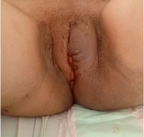
Apart from the hemoglobin level of 4.3 grams/deciliter, the preoperative workup was unremarkable. The intraoperative findings were: a hematoma of 1100 milliliters located between the superficial and deep planes of the perineum and active bleeding from the superficial and deep perineal arteries. After evacuating the hematoma, we successfully performed several X shaped hemostatic sutures using resorbable braided polyglactin thread 0 suture. The residual cavity was then easily lined. Finally, the homolateral vaginal tear was sutured. No bleeding was noted afterwards in the immediate postoperative surveillance (Figure 3).
The postoperative course was unremarkable with good involution of the vulvar hematoma and disappearance of the discharge. The patient was discharged with her child at 2 days postpartum. Three weeks later the vulvovaginal anatomy was macroscopically normal.

Discussion
Vulvar hematoma is a clinical entity that occurs mainly in the obstetrical context. Outside of pregnancy and postpartum, these diverse and even unusual circumstances of onset have been reported: straddle fall trauma and post-coital [8-10]. The frequency of puerperal vulvar hematoma varies from small (1 case per 700 deliveries) to large (1 case per 4000 deliveries) [6]. The risk factors for this complication recognized in the literature are: primiparity, instrumental fetal extraction, pre-eclampsia, twin pregnancies, macrosomia and vulvovaginal varices [6]. In our patient, we found the use of the vacuum cup to assist fetal extraction.
As reported in the literature, the clinical picture in our patient was dominated by the occurrence of pain and swelling, usually unilateral, a few moments or immediately after delivery [6,11]. Homolateral vaginal tearing and uterine atony were associated, which aggravated the ensuing hemorrhagic shock. The literature reports several topographic forms of puerperal hematoma: vulvar hematoma, vaginal hematoma, vulvovaginal hematoma, and pelvic-genital or subperitoneal hematoma [6]. Given the significant development of the vascularization of the perineal area during pregnancy and especially during delivery, the expansive vulvar hematoma is often complicated by hemorrhagic shock which can be deadly [11,12].
Management remains primarily surgical with the use of X-stitches with absorbable thread followed by padding to ensure hemostasis [13,14]. Anesthesia can be either local or general [14]. The evolution is most often favorable and depends on the promptness of diagnosis and management [14].
Conclusion
Postpartum vulvar hematoma is an uncommon but serious postpartum complication. It can be associated with a state of shock which can threaten the vital prognosis of the parturient. Practitioners should be aware that the outcome depends on the promptness of diagnosis and management.
Declarations
Guarantor of submission: The corresponding author is the guarantor of submission.
Acknowledgements: None.
Funding: There are no funding sources to be declared.
Availability of data and materials: Supporting material is available if further analysis is needed.
Competing interests: The authors declare that they have no competing interests.
Consent for publication: Written informed consent was obtained from the patient for publication of this case report and any accompanying images. A copy of the written consent is available for review by the Editor-in-Chief of this journal.
Ethics approval and consent to participate: Ethics approval has been obtained to proceed with the current study. Written informed consent was obtained from the patient for participation in this publication.
References
- Abdul Kadir R, Mc Lintock C, Ducloy AS, El-Refaey H, England A, et al. Evaluation and management of postpartum hemorrhage: Consensus from an international expert panel. Transfusion. 2014; 54: 1756-1768.
- Talib S, Slaoui A, Mahtate M, et al. Massive Vulvar Edema Complicating Severe Preeclampsia: A Case Report. Arch Surg Clin Case Rep. 2020; 3: 144.
- World Health Organisation Department of Reproductive Health and Research. Maternal mortality in 2000: estimates developed by WHO, UNICEF, and UNFPA. 2000.
- Slaoui A, Talib S, Nah A, et al. Placenta accreta in the department of gynaecology and obstetrics in Rabat, Morocco: Case series and review of the literature. Pan Afr Med J. 2019; 33: 86.
- Riethmuller D, Pequegnot-Jeannin C, Rabenja CA, Koeberle P, Schaal JP, et al. A rare cause of postpartum hemorrhage: A genital thrombus. J Gynecol Obstet Biol Reprod (Paris). 1997; 26: 154 -158.
- Bienstman Pailleux J, Huissoud C, Dubernard G, Rudigoz RC. Priseen charge des hematomas Puepéraux. Journal de Gynécologie Obstétrique et Biologie de la Reproduction. 2009; 38: 203–208.
- Slaoui A, Slaoui A, Zeraidi N, Lakhdar A, Rhrab B, Kharbach A, Baydada A. Benckiser’s hemorrhage: about an uncommon case report, Int J Surg Case Rep. 2022; 95: 107128. https://doi.org/10.1016/j.ijscr.2022.107128.
- Virgili A, Bianchi A, Mollica G, Corazza M. Serious hematoma of the vulva from a bicycle accident: a case report. J Reprod Med. 2000; 45: 662-664.
- Tjek P, Essiben F, Moluh I, Tebeu PM, Fomulu JN. Hématome vulvaire massif gauche post coïtal. Health Sci Dis. 2013; 14: 3.
- Kehila M, Khedher SB, Zeghal D, Mahjoub S. Prise en charge conservatrice des hématomes puerpéraux de gros volume: A propos de 3 cas. The Pan African Medical Journal. 2013; 16: 9.
- Alirezaei S, Vatanchi A, Pourali L, Aminzadeh B, Latifnejad Roudsari R. Mortality of a postpartum woman presented with massive vulvar edema in association with Covid-19: A case report with clinical and radiological findings. BMC Infect Dis. 2021; 21: 678.
- Tilahun T, Wakgari A, Legesse A, Oljira R. Postpartum spontaneous vulvar hematoma as a cause of maternal near miss: A case report and review of the literature. J Med Case Rep. 2022; 16: 85.
- Slaoui A, Slaoui A, Himmi Y, Mouftah B, Mamad A, et al. Transvaginal and transobturator autologous vaginal tape cystocele treatment: About an uncommon case. Int J Surg Case Rep. 2022; 95: 107198.
- Tseng JY, Lin IC, Lin JH, Chang CM, Chao WT, et al. Optimal approach for management of postpartum vulva hematoma: Report of three cases. Taiwan J Obstet Gynecol. 2020; 59: 780-783.