
Journal of Clinical Images and Medical Case Reports
ISSN 2766-7820
Clinical Image - Open Access, Volume 3
Bronchial atresia presenting with two bronchoceles
Inês Barreto1*ⴕ; Inês Spencer1ⴕ; Francisca Godinho Oliveira1; João Rodrigues Inácio2; Richard Staats1; Cristina Bárbara1
1Department of Pulmonology, North Lisbon University Hospital Centre, Portugal.
2Department of Radiology, North Lisbon University Hospital Centre, Portugal.
ⴕEqual Contribution.
*Corresponding Author : Inês Barreto
Department of Pulmonology, North Lisbon University Centre, Portugal.
Email: inesscbarreto@gmail.com
Received : Aug 19, 2022
Accepted : Sep 07, 2022
Published : Sep 14, 2022
Archived : www.jcimcr.org
Copyright : © Barreto I (2022).
Keywords: Bronchial atresia; Bronchocele; Mucocele; Congenital abnormality; Segmental hyperinflation.
Citation: Barreto I, Spencer I, Oliveira FG, Inácio JR, Staats R, et al. Bronchial atresia presenting with two bronchoceles. J Clin Images Med Case Rep. 2022; 3(9): 2051.
Background and case description
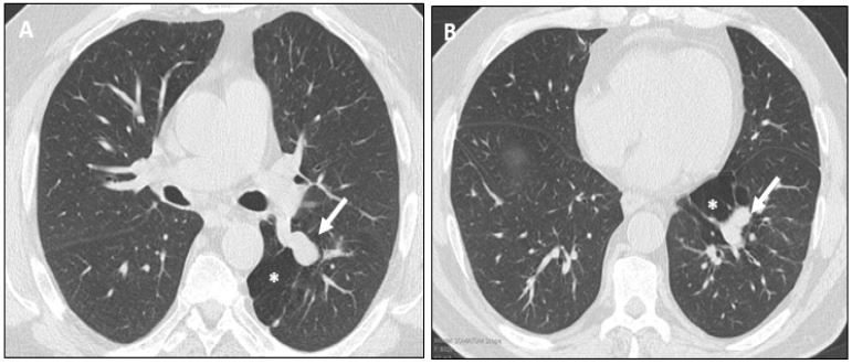
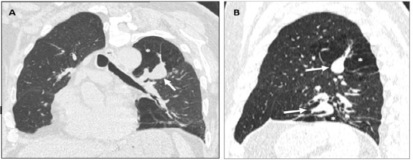
Bronchial atresia is a rare congenital lung abnormality characterized by the focal obliteration of the bronchial lumen, on a lobar, segmental or subsegmental level, usually associated with peripheral mucus impaction (bronchocele or mucocele) [1,2]. It presents with adjacent segmental or subsegmental hyperinflation, and reduction of vascularization of the involved segment or lobe.
A 66-year-old Caucasian man, former smoker (40 pack-years), with past medical history of arterial hypertension, type 2 diabetes mellitus, dyslipidemia, and obstructive sleep apnea was evaluated due to snoring and insomnia. The physical examination and laboratory tests were unremarkable. The pulmonary function tests presented no abnormalities.
Chest Computed Tomography (CT) identified at the superior and medial basal segments of the left lower lobe, hyperinflation and hyperlucent lung parenchyma, with tubular-shaped opacities with no communication with the adjacent bronchial tree representing bronchoceles. The findings were consistent with bronchial atresia presenting with two segmental bronchoceles of the left lower lobe.
Bronchial atresia is a developmental lung abnormality usually presenting as an incidental finding on imaging. The presentation with two bronchoceles is uncommon. Although it may be associated with recurrent infections in 20% of the cases, most of the patients are young, asymptomatic and have no abnormalities on physical examination or laboratory tests. Chest CT is the preferred modality for diagnosing bronchial atresia [1]. Treatment is conservative in asymptomatic patients. When there is suspicion of malignancy as the cause of obstruction or complications secondary to the atretic bronchus, surgical resection may be performed [1].
Acknowledgements: Inês Barreto and Inês Spencer have contributed equally.
References
- Gipson Matthew G, Cummings Kristopher W, Hurth Kyle M. Bronchial Atresia. Radiographics. 2009; 29: 1531–1535.
- Berrocal T, Madrid C, Novo S, et al. Congenital anomalies of the tracheobronchial tree, lung, and mediastinum: Embryology, radiology, and pathology. Radiographics. 2004; 24: e17.