
Journal of Clinical Images and Medical Case Reports
ISSN 2766-7820
Case Report - Open Access, Volume 3
Spontaneous enterocutaneous fistula secondary to femoral hernia: A case report
Akhil Raj Anumolu*; Kiran Kumar Miriyam
Andhra Medical College, Visakhapatnam, India.
*Corresponding Author : Akhil Raj Anumolu
Andhra Medical College, Visakhapatnam, India.
Phone: +91-833-186-0100;
Email: akhilrajanumolu@yahoo.com
Received : Aug 22, 2022
Accepted : Sep 09, 2022
Published : Sep 16, 2022
Archived : www.jcimcr.org
Copyright : © Anumolu AR (2022).
Abstract
Background: Femoral hernias account for 10% of all groin hernias. Females have a 4:1 incidence of abdominal hernias, while males have a 5% incidence. A spontaneous enterocutaneous fistula is rare due to a small bowel loop perforation in an incarcerated femoral hernia. Patients’ ignorance and late presentation cause these fistulas.
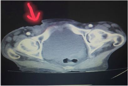
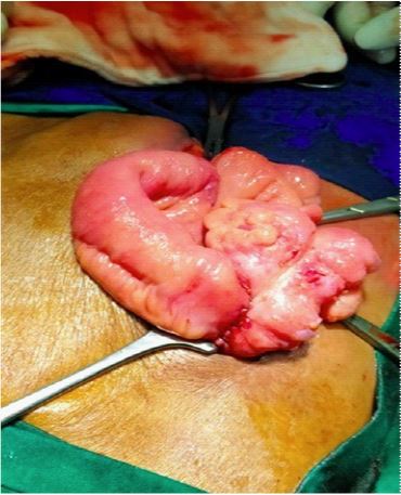
Case: A 65-year-old female presented to an emergency department of a public hospital with a two-year-old right groin swelling. A Contrast-Enhanced Computed Tomography (CECT) scan revealed a defect in the anterior abdominal wall in the right groin through which the small bowel loop protruded. It was 20 cm from the ileocecal junction. For the femoral hernia, Cooper ligament repair was used. Ileo ascending colon anastomosis was performed after gangrenous segment resection. Finally, the previously existing skin defect was left to heal.
Conclusion: This case report discusses the presentation of spontaneous enterocutaneous fistula due to femoral hernia, as very little literature describes this phenomenon.
Citation: Anumolu AR, Miriyam KK. Spontaneous enterocutaneous fistula secondary to femoral hernia: A case report. J Clin Images Med Case Rep. 2022; 3(9): 2056.
Introduction
Femoral hernia cases constitute 10% of all groin hernias. They are common in females with a female to male ratio of 4:1 and a 20% incidence amongst all abdominal hernias in females and 5% in males [1]. Groin hernias become complicated due to chronicity or sudden precipitating episodes. Of all groin hernias, the femoral hernia is relatively uncommon and is more prone to complications like incarceration and strangulation as it passes through a narrow femoral ring. Enterocutaneous fistula commonly occurs postoperatively in patients undergoing bowel surgeries [2]. Almost 85% of cases occur postoperatively with an increased incidence in the emergency surgical setting [3]. Other common causes include radiation, inflammatory bowel disease, diverticulitis, and appendicitis [3]. Spontaneous enterocutaneous fistula due to perforation of a small bowel loop in incarcerated femoral hernia is rare and only a few cases have been reported. Such fistulae often arise due to ignorance of patients and delayed presentation [4].
Case presentation
A 65-year-old female presented to the emergency department in a public hospital for urgency, with a history of a neglected swelling in the right groin for the past two years. She had no history of nausea, vomiting, fever, colicky abdominal pain, or abdominal distention. However, four days prior to her visit, she noticed that the skin around the swelling had suddenly become red and painful after an episode of strenuous work, and subsequently, the swelling ruptured pouring out feculent content. She had chief complaints of pain in the right groin for the past four days and feculent discharge from the swelling for the past three days. Upon examination, the vitals and general condition of the patient were stable and there were no signs of peritonitis. She did not have any features of obstruction, albeit there was an enterocutaneous fistula in the right groin region. No swellings were present in the left groin or in the umbilical region. The patient did not have evidence of sepsis.
Discussion
In most cases of femoral hernia, they contain a loop of bowel herniating through them, which are titularly termed Richter’s hernia (partial enterocele). Such cases rapidly progress to gangrene as the constricting ring exerts pressure on the wall of the bowel, obstructing blood flow. Signs and symptoms of intestinal obstruction are present only in cases where more than two-thirds of the bowel wall circumference is involved [5]. In the case of our patient, she had a complete loop of bowel in the femoral ring in contrast with the usual Ritcher’s type hernia usually reported.
An Enterocutaneous Fistula (ECF) forms in the presence of an abnormal communication between the gastrointestinal tract and the skin (or wound). ECFs most commonly follow a visceral surgery (i.e. iatrogenic) [6,7]. They have a higher incidence postoperatively with only one-fourth of cases occurring spontaneously, most frequently in the setting of inflammatory bowel diseases like Crohn’s [7]. Other common causes of spontaneous ECFs are malignancies, post-radiation therapy for malignancies, diverticulitis, appendicitis, mesenteric ischemia, tuberculosis, and strangulation of bowel loops [5,8]. The evolution of an incarcerated hernia to an ECF results in bowel decompression and is associated with an increased incidence of sepsis and mortality, especially with high-output fistulae (>500 mL/24 h) [3].
They have a high propensity for strangulation and incarceration with an increasing probability of complications as time passes. They commonly present with obstruction and perforation of the incarcerated ileal loop leading to the formation of ECFs in these patients is a rare phenomenon, as seen in our patient [6]. Additionally, our patient’s case was similar to a Ritcher’s type of hernia as only part of the bowel wall was trapped in the hernial sac even though a complete loop had herniated, which led to the formation of an ECF [9].
The common acronym SNAP is used as a guideline to describe ECF care protocol and has been proven effective in the treatment of such cases [7]. It involves initial resuscitation, electrolyte repletion, identification, and treatment of sepsis followed by a nutritional assessment. In patients who are well-nourished and with effectively controlled fistula effluent, this is followed by imaging to view the involved anatomical structures. The final step is the closure of the fistula through a surgical procedure [7]. Our patient was managed in accordance with the SNAP guideline which led to steady improvement and an absence of complications.
Our case of spontaneous ECF in the setting of a complicated femoral hernia was due to late presentation, negligence, and poor socioeconomic status of the patient. Most patients in our setup belong to poor settings, lack the appropriate knowledge required for timely check-ups, and neglect their health until critical levels or emergencies. Delayed presentation is the major cause of increased hospital stay, morbidity, and mortality. These factors play an important role in the formation of such fistulae even with the technological and innovative advances in the medical field. However, management constituting adherence to protocols like the SNAP guideline is essential for delayed presentations to avoid further complications. After correcting electrolyte abnormalities, patients should be subjected to a defini tive surgical procedure, as early as possible.
References
- Nikolopoulos I, Oderuth E, Ntakomyti E, Kald B. Intestinal Obstruction due to Bilateral Strangulated Femoral Hernias. Case Rep Surg. 2014; 2014: 195736.
- Sistla SC, Reddy R, Dharanipragada K, Jagdish S. Enterocutaneous fistula due to mesh fixation in the repair of lateral incisional hernia: a case report. Cases J. 2008; 1: 370.
- Berry SM, Fischer JE. Classification and pathophysiology of enterocutaneous fistulas. SurgClin North Am. 1996; 76: 1009-1018.
- Bostanci ME, Özel İ, Bozkurt B, Soylu S, Turan M, et al. Spontaneous enterocutaneous fistula: A rare presentation of incarcerated femoral hernia. Eurasian Journal of Emergency Medicine. 2015; 14: 199.
- Ahi KS, Moudgil A, Aggarwal K, Sharma C, Singh K. A rare case of spontaneous inguinal faecal fistula as a complication of incarcerated Richter’s hernia with brief review of literature. BMC Surg. 2015; 15: 67.
- Kumar A, Pahwa HS, Pandey A, Kumar S. Spontaneous enterocutaneous fistula due to femoral hernia. BMJ Case Rep. 2012; 2012: bcr2012006939.
- Gribovskaja Rupp I, Melton GB. Enterocutaneous Fistula: Proven Strategies and Updates. Clin Colon Rectal Surg. 2016; 29: 130-137.
- Shah M, Wani MA. Spontaneous Tubercular Enterocutaneous Fistula. Saudi J Med Med Sci. 2017; 5: 275-277.
- Skandalakis PN, Zoras O, Skandalakis JE, Mirilas P. Richter hernia: Surgical anatomy and technique of repair. Am Surg. 2006; 72: 180-184.