
Journal of Clinical Images and Medical Case Reports
ISSN 2766-7820
Clinical Image - Open Access, Volume 3
Pyogenic granuloma of the glans penis after
angioembolisation procedure
Deepak Yumnam*; Anmol Batra; Rikita Paonam; Avita Dhiman
Department of Dermatology, Venereology, and Leprology, All India Institute of Medical Sciences, Rishikesh, Uttarakhand 249203, India.
*Corresponding Author : Deepak Yumnam
Academic Junior Resident, Department of Dermatology, Venereology and Leprology, All India Institute of Medical Sciences, Veerbhadra Marg, Rishikesh, Uttarakhand (249203), India.
Tel: +91-763-090-6798;
Email: deepakyumnam@gmail.com
Received : Aug 26, 2022
Accepted : Sep 13, 2022
Published : Sep 20, 2022
Archived : www.jcimcr.org
Copyright : © Yumnam D (2022).
Citation: Yumnam D, Batra A, Paonam R, Dhiman A. Pyogenic granuloma of the glans penis after angioembolisation procedure. J Clin Images Med Case Rep. 2022; 3(9): 2063.
Case report
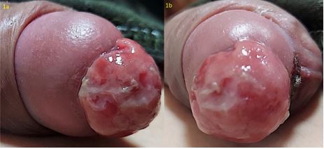
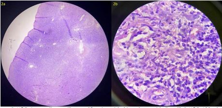
A 8-year-old male child was referred to the Dermatologic clinic for a protruding growth on the glans penis that had appeared and started growing rapidly over a period of past 7 days since the child underwent the angioembolisation procedure for Penile Arteriovenous (AV) malformation. There was a history of intermittent bleeding from the well-established lesion. Clinical examination revealed a single, well-defined, rounded, sessile, fleshy red, vascular, polypoidal growth of size 3.5 x 3 cm present circumferentially around the urethral opening on the glans penis (Figure 1). The patient’s clinical course and a physical examination for glans penis lesion was suggestive of a Pyogenic Granuloma (PG). Histopathological examination of the mucosal biopsy from the lesion confirmed the diagnosis showing proliferating vessels surrounded by a mixed inflammatory infiltrate comprising lymphocytes, plasma cells, neutrophils, and eosinophils. After the confirmation of the diagnosis, the lesion was completely excised under local anaesthesia. During the postoperative follow-up, there was no recurrence six months after the surgery.
Pyogenic granuloma, a misnomer, also known as lobular capillary hemangiomas are acquired, benign, vascular proliferations arising from the skin and mucous membranes and not due to infection. There have been a variety of proposed causes of PG and is considered as reactive phenomenon. Recent minor trauma accounts for 7% of presentations [1]. Little is known regarding the occurrence of PG in the glans penis. PG on the glans penis is rare in pediatric age group, only few cases have been reported [2,3]. It may also develop after circumcision. PG often develops an eroded surface, with subsequent bleeding which can be profuse. Early treatment is required as it may grow rapidly and often undergoes ulceration. Surgeons should be careful while doing the procedure including the instrumentation and even catheterisation to minimize trauma. Simple curettage with electrocautery is usually curative in small lesion. Other options include excision, laser surgery (carbon dioxide or pulsed-dye laser), and cryotherapy. Imiquimod and timolol have been suggested as effective topical treatment options [1].
References
- Tritton SM, Smith S, Wong LC, et al. Pyogenic granuloma in ten children treated with topical imiquimod. Pediatr Dermatol. 2009; 26: 269-272.
- Eickhorst KM, Nurzia MJ, Barone JG. Pediatric pyogenic granuloma of the glans penis. Urology. 2003; 61: 644.
- Spinelli C, Di Giacomo M, Bertocchini A, Loggini B, Pingitore R, et al. Multiple pyogenic granuloma of the penis in a four-year-old child: A case report. Cases J. 2009; 2: 7831.