
Journal of Clinical Images and Medical Case Reports
ISSN 2766-7820
Case Report - Open Access, Volume 3
Native aortic valve thrombosis in a patient with chronic thromboembolic pulmonary hypertension
Selbi Meiramova1*; Timur Lesbekov1; Aigerim Kuzhakhmetova1; Aidyn Kuanyshbek2; Aigerim Kunakbaieva1
1Cardiac Surgery Department, National Research Cardiac Surgery Center, Turan, 38, 010000 Nur-Sultan, Kazakhstan.
2Anesthesiology and Intensive Care Department, National Research Cardiac Surgery Center, Turan, 38, 010000 Nur-Sultan, Kazakhstan.
*Corresponding Author : Selbi Meiramova
National Research Cardiac Surgery Center, Department of Cardiac Surgery, 38 Turan street, Z05G9F9 Astana, Kazakhstan.
Email: selvi_m.d@mail.ru
Received : Aug 27, 2022
Accepted : Sep 21, 2022
Published : Sep 28, 2022
Archived : www.jcimcr.org
Copyright : © Meiramova S (2022).
Abstract
Spontaneous native aortic valve thrombosis is an unusual cause of coronary insufficiency and more common in patients with underlying coagulopathies, such as antifospholipid syndrome, valve disease or bacterial endocarditis. However in this case we want to demonstrate a case that doesn’t fit above mentioned criteria. Even more, a rare condition of spontaneous native aortic valve thrombosis is combined with Chronic Thromboembolic Pulmonary Hyperthension (CTEPH).
Keywords: Spontaneous native aortic valve thrombosis; Chronic thrmboembolic pulmonary hypertension.
Citation: Meiramova S, Lesbekov T, Kuzhakhmetova A, Kuanyshbek A, Kunakbaieva A, et al. Native aortic valve thrombosis in a patient with chronic thromboembolic pulmonary hypertension. J Clin Images Med Case Rep. 2022; 3(9): 2077.
Introduction
Native aortic valve thrombosis is an uncommon condition with a high (around 20%) in-hospital mortality rate [1]. On the other hand CTEPH is a rare condition that develops as a result of pulmonary embolism in a range of 0,1 to 4,0% of cases [2]. In this paper we present the first known case of two conditions combined.
Case report
A 47-years old female was hospitalized on 13/08/2021 for elective Pulmonary Thrombendarterectomy (PTE) due to chronic thromboembolic pulmonary hypertension. According to the anamnesis, diagnosis was verified 6 month prior to hospitalization with underlying cause being acute lower extremities deep vein thrombosis. Therapy was prescribed according to the protocol with New Oral Anticoagulants (NOAC) among other medications.
Additionally, on 10/08/2021 the patient underwent resection of submucous myoma with suturing of the myoma bed, which was the cause of interruption of anticoagulant therapy for 3 days.
Laboratory testing revealed no coagulopathies; ECG showed sinus rhythm with HR 75 bpm, right ventricular overload and partial right bundle branch block: Changes on TTE correlated with the main diagnosis (right heart enlargement, pulmonary artery dilatation, secondary tricuspid regurgitation; EF 67,07%, EDV LV 85 ml). Prior angiography showed intact coronary arteries.
Comorbidity included type 2 diabetes and anemia.
16/08/2021 at 14:30 patient presented with acute onset chest pain, accompanied with shortness of breath, hypotension up to 70/40 mmHg. ECG showed ST segment elevation in leads V1-V6, laboratory test revealed troponin levels of 7,9 ng/dl. Furthermore, patient had paroxysms of ventricular fibrillation with hemodynamic instability.
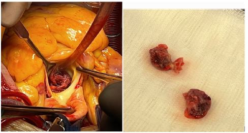
With signs of cardiogenic shock patient was taken to the operation room. TEE revealed floating thrombotic masses in noncoronary and left coronary sinus obstructing respective artery (Figure 1), hypokinesis of anterior, septal and apical segments of left ventricle, acute (ischemic) mitral regurgitation.
CPB was initiated (ascending aorta – superior and inferior vena cava). White thrombotic masses in the form of casts were easily removed from noncoronary and left coronary sinuses (Figure 2). Valve itself was tricuspid, leaflets were thin and the surface was smooth, hence valve replacement was not required. CABG (SVG to OM and LAD) and thrombecthomy from left branch of pulmonary artery were performed. Due to acute ischemic nature of mitral regurgitation, considering acute heart failure because of myocardial infarction, volume of surgery, the decision was made to finish the procedure with central V-A ECMO (right atrium – ascending aorta).
Here-in-after patient was treated in ICU. Attempt to wean patient off ECMO on 18/08/2021 was unsuccessful with hypotension and lactate acidosis in response to reducing the flow on ECMO. In addition severe mitral regurgitation persisted.
However, 18/08/2021 in the ICU patient was extubated and activated. Laboratory tests showed typical peaks and decreases of troponin levels, otherwise uneventful.
24/08/2021 patient was successfully weaned of ECMO. Intraoperatively: venous grafts are potent, no signs of MR. She was extubated the same day and transferred to the surgical unit on 25/08/2021.
Further postoperative period was uneventful, control TTE showed LV EF 45%, no signs of mitral insufficiency, ECG revealed negative T waves in leads V2-V6.
Coronary angiography performed 4 month after the surgery confirmed grafts’ potency.
Heart catheterization with pulmonary angiography 4 month later revealed no signs of rethrombosis or residual pulmonary hypertension.

Discussion
Coronary artery embolism is a rare nonatherosclerotic cause of ST-elevation myocardial infarction and varies from 4 to 13% according to autopsy or angiographic data [3]. Native aortic valve thrombosis is an extremely rare condition, with STEMI incidence of about 36% [3].
This case got our attention because due to CTEPH patient was prior examined on all main predisposing hypercoagulable conditions, but no abnormality was found. Moreover, TTE performed 4 days prior ischemic events showed no signs of heart chambers and valves thrombosis, although in the literature hypercoagulable states are the main cause of native valve thrombosis [3].
Another risk factors include degenerative or congenital valve disease [4], prior catheter intervention [5] or bacterial endocarditis [6], none of which is fit for our case.
Hystopathological examination confirmed excised mass as organized thrombus formation, which leads us to the conclusion of idiopathic native aortic valve thrombosis.
Conclusion
To the best of our knowledge, this is the first reported case of native aortic valve thrombosis complicated with CS in a patient with confirmed CTEPH. Perhaps although all of the standard coagulopathies were excluded, diagnostic search must not be stopped. Additionally, patients with known venous thrombosis must be thoroughly examined for arterial one.
In addition, the patient has provided permission to publish these features of her case, and the identity of the patient has been protected.
References
- Alajaji W, Hornick JM, Malek E, Klein AL, et al. The Characteristics and Outcomes of Native Aortic Valve Thrombosis: A Systematic Review. J Am Coll Cardiol. 2021; 78: 811-824.
- John DL Brookes, Crystal Li, Sally TW Chung, Elizabeth M Brookes, Michael L, et al. Williams, Nicholas McNamara, Sofia Martin-Suarez, Antonio Loforte. Pulmonary thromboendarterectomy for chronic thromboembolic pulmonary hypertension: A systematic review. Ann Cardiothorac Surg 2022; 11: 68-81. https://dx.doi.org/10.21037/acs-2021-pte-25
- Popovic B, Agrinier N, Bouchahda N, Pinelli S, Maigrat CH, et al. Coronary Embolism Among ST-Segment-Elevation Myocardial Infarction Patients: Mechanisms and Management. Circ Cardiovasc Interv. 2018; 11: e005587.
- Wan S, DeSmet JM, Vincent JL, LeClerc JL. Thrombus formation on a calcific and severely stenotic bicuspid aortic valve. Ann Thorac Surg. 1997; 64: 535-536.
- Rait MH, Schwaegler B, Pearlman AS, Poole JE, Bardy GH, Dolack GL, et al. Development of an aortic valve mass after radiofrequency catheter ablation. PACE. 1993; 16: 2064-2066.
- Castillo JC, Anguita MP, Torres F, Siles JR, Mesa D, Vallés F, et al. Risk factors associated with endocarditis without underlying heart disease. Rev Esp Cardiol. 2002; 55: 304–307.