Journal of Clinical Images and Medical Case Reports
ISSN 2766-7820
Clinical Image - Open Access, Volume 3
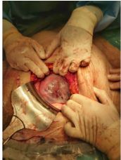
Couvelaire uterus after external cephalic version
*Corresponding Author : María del MR Arroyo
Arcipreste de Hita Nº3 1ºB Guadalajara 19001,
Spain.
Tel: 655-854-214;
Email: mar_gotor@hotmail.com
Received : Aug 26, 2022
Accepted : Sep 22, 2022
Published : Sep 29, 2022
Archived : www.jcimcr.org
Copyright : © Arroyo MMR (2022).
Citation: Arroyo MMR. Couvelaire uterus after external cephalic version. J Clin Images Med Case Rep. 2022; 3(9): 2078.
Description
A 30-years-old primiparous woman with 37 weeks of amenorrhoea was admitted to the Obstetric ward with symptoms of ruptured membranes and abdominal pain. The day before she was subjected to an unsuccessful external cephalic version. Transabdominal ultrasonography revealed a breech presentation. The placenta appeared normal. The Cardiotocograph (CTG) showed a nonreassuring pattern with deep variable decelerations. An emergency cesarean section was performed. Intraoperatively, the uterus showed a large intramyometrial hematoma coinciding with the operator`s side of the external cephalic version the previous day. A retroplacental hematoma was evidenced after hysterotomy. The diagnosis of placental abruption and Couvelaire`s uterus was established. The newborn`s 1-minute and 5-minute Apgar scores were 8 and 9, respectively. The postoperative period was good and mother and son are progressing favourably.