
Journal of Clinical Images and Medical Case Reports
ISSN 2766-7820
Case Report - Open Access, Volume 3
Bilateral ocular ischemic syndrome following treatment
for laryngeal carcinoma
Archana Nair; Edward Chaum*
Vanderbilt University Medical Center, Nashville, TN, USA.
*Corresponding Author : Edward Chaum
Vanderbilt Eye Institute, Vanderbilt University Medical Center, Nashville TN, 37232, USA.
Email: edward.chaum@vumc.org
Received : Aug 15, 2022
Accepted : Sep 28, 2022
Published : Oct 05, 2022
Archived : www.jcimcr.org
Copyright : © Chaum E (2022).
Citation: Nair A, Chaum E. Bilateral ocular ischemic syndrome following treatment for laryngeal carcinoma. J Clin Images Med Case Rep. 2022; 3(10): 2087.
Description
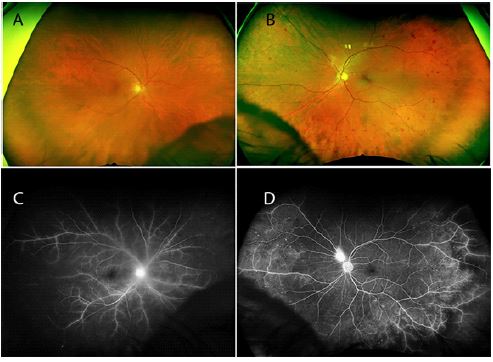
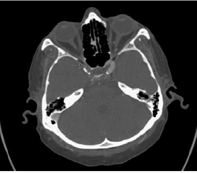
A 72-year-old male with a history of atherosclerosis and radiation therapy for laryngeal cancer presented with complaints of decreased vision. The exam of the right eye was notable for neovascularization of the iris and significant vaso-occlusive disease of the retina, with non-perfusion of the peripheral retina (Figure 1A). Classical large dot-blot hemorrhages were present in the mid-periphery of left retina consistent with the diagnosis of bilateral Ocular Ischemia Syndrome (OIS) (Figure 1B). Fluorescein angiography demonstrated more severe OIS of the right retina, with global peripheral capillary drop out and optic disc and diffuse vascular leakage (Figure 1C). Far peripheral capillary drop out was seen in the left eye with leaking neovascular vessels near the disc (Figure 1D). CT angiography showed arterial occlusive disease with impairment of carotid artery perfusion of the clinoid segment of the right internal carotid artery on axial CT angiogram, mirroring the severity of retinal ischemia between the two eyes (Figure 2).
Ocular Ischemic Syndrome (OIS) was initially described as a spectrum of ocular findings characterized by retinal hemorrhages and dilated retinal veins [1], and is commonly associated with neovascularization of the iris, anterior segment inflammation, and other ocular signs and symptoms attributable to significant carotid artery obstruction. The condition occurs in about 2100 patients per year in the United States (M:F ratio is 2:1) and is bilateral in ~20% of patients. Iris neovascularization is present in two-thirds of eyes at the time of presentation. OIS appears to develop when carotid artery blood flow obstruction reaches ~90%.
The major risk factor for OIS in most patients is atherosclerosis (which was present in our patient) but the condition, in particular bilateral presentation, has been reported in systemic arterial inflammatory diseases including Takayasu arteritis and giant cell arteritis [2]. It has also been seen as a late complication of radiotherapy for nasopharyngeal carcinoma [3]. To our knowledge this is the first reported case of bilateral OIS associated with radiation treatment for laryngeal carcinoma, which may have contributed to his disease and its severity in both eyes [4].
References
- Kearns TP, Hollenhorst RW. Venous-stasis retinopathy of occlusive disease of the carotid artery. Mayo Clin Proc. 1963; 38: 304–312.
- Mendrinos E, Machinie TG, Pournaras CJ. Ocular Ischemic Syndrome. Surv Ophthalmol. 2010; 55: 2-34.
- Tang Y, Luo D, Peng W, Huang F, Peng Y, et al. Ocular ischemic syndrome secondary to carotid artery occlusion as a late complication of radiotherapy of nasopharyngeal carcinoma. J Neuroophthalmol. 2010; 30: 315-320.
- Avitia S, Hamilton J, Osborne RF. Radiation-induced carotid artery stenosis. Ear Nose Throat J. 2006; 85: 158.