
Journal of Clinical Images and Medical Case Reports
ISSN 2766-7820
Review Article - Open Access, Volume 3
Could machine learning approach improve diagnosis accuracy for malignancy in imaging examinations? A mini-review
Chao Yang1; Jianhui Xu1,2; Yingshi Zhang3*
1Department of Ethnic Culture and Vocational Education, Liaoning National Normal College, Shenyang 110031, China.
2Faculty of Ocean Engineering Technology and Informatics, Universiti Malaysia Terengganu, Terengganu 21030, Malaysia.
3Department of Clinical Pharmacy, Shenyang Pharmaceutical University, Shenyang 110016, China.
*Corresponding Author : Yingshi Zhang
Department of Clinical Pharmacy, Shenyang Pharmaceutical University, Shenyang, 110016 China.
Email: zhangyingshi526@163.com
Received : Sep 06, 2022
Accepted : Oct 04, 2022
Published : Oct 11, 2022
Archived : www.jcimcr.org
Copyright : © Zhang Y (2022).
Abstract
The early diagnosis of malignancy by ultrasound, Computed Tomography (CT), and Magnetic Resonance Imaging (MRI) is still limited. Therefore, the diagnostic efficacy could be improved from Machine Learning (ML), which mainly based on deep learning and convolutional neural networks. In this work, we reviewed ML-based imaging examinations ultrasound, CT and MRI could evaluate early diagnosis value of small size tumor. Besides, ultrasound could distinguish primary tumors from metastatic tumors, CT could also play a role in tumor risk classification, and MRI could also predict the pathological grade and enzyme mutation status of malignancy, which can be used to predict early survival and guide clinical decision-making. Therefore, we believe that ML could be added to improve the accuracy of diagnosis in patients with suspected tumor imaging examinations.
Citation: Yang C, Xu J, Zhang Y. Could machine learning approach improve diagnosis accuracy for malignancy in imaging examinations? A mini-review. J Clin Images Med Case Rep. 2022; 3(10): 2095.
Introduction
Malignancy is a huge social and health burden for human beings [1,2]. Malignancy has an feature of insidious onset in the early stage, develops rapidly in the middle period, and is likely to metastasize and relapse in the late period. Besides, cancer is insensitive to radiotherapy and chemotherapy. Once molecular targeted agents resistance occurs, there are no molecular agents available, which causing irreparable losses such as patient death [3-6]. Thus, it is especially important to diagnose malignant tumors at an early stage, and there are limitations in the diagnostic accuracy of ultrasonography, Computed Tomography (CT) and Magnetic Resonance Imaging (MRI) [7-9]. Moreover, some tumor markers such as AFP, DKK-1, OPN, etc., which are currently used in the clinic are still limited in their diagnostic efficacy in distinguishing tumors [10,11]. Therefore, we need to find an effective way to enhance the sensitivity of existing cancer diagnosis, such as Machine Learning (ML) to enhance imaging examination.
ML-based approaches provide clinical application in the accumulation extraction of key imaging characteristics and measures, including image classification and lesion segmentation, which show that ML is used in cancer detection to stratify high risk individuals for cancer prediction. This is due to the fact that ML-based approaches provide a wide range of automated tools for performing radiomics for the purpose of detecting and quantifying tissue properties [12-14]. The implementation method is though highly complex Artificial Intelligence (AI) models, which are based on a series of interconnected mathematical equations, have been proposed for the analysis of complicated, high dimensional health data, such as Deep Learning (DL) include Convolutional Neural Networks (CNNs) and Recurrent Neural Networks (RNNs) [15-18]. In recent years, significant progress has been made in applying ML to interpreting medical imaging, mainly because of DL algorithms utilizing CNN. However, there is no systematic review on whether ML can increase the diagnostic efficacy of imaging examinations.
ML approach for malignancy diagnosis using ultrasound
Ultrasound is one of the most popular imaging methods in clinic due to its low cost and easy operation, which can offer real time images [19]. The aim of Wu T is to assess the potential of ML in diagnosing Triple Negative (TN) breast cancer by means of quantitative ultrasound. A retrospective analysis was made on the ultrasound and clinical features of 140 patients with surgical confirmation of breast cancer in order to diagnose TN and Non TN (NTN) subtypes. Among 12 gray scale and Doppler characteristics, 8 showed significant differences between TN and NTN subtypes (p < 0.05). The Area below ROC (AUC) was 0.85 and 0.65 for Gray Scale (GS) and Color Doppler (CD). The AUC was raised to 0.88 when combined with GS and CD characteristics. The sensitivity was 86.96%, and the specificity was 82.91%. Finally, we conclude that the TN and NTN sub-types can be distinguished from each other by means of ML, which is superior to the diagnosis by means of standard visual evaluation [20].
Moreover, the aim of this research is to detect the presence of cervical cancer by means of a cervicogram. They employed a deep learning framework, ResNet-50, to classify 4,119 cervicogram images as positive or negative for cervical carcinoma, with a rectangle with a section of the vaginal wall removed. ML models were used to extract the data from more than 300 images. The ResNet-50 model improved by 0.15 (p < 0.05) compared with the mean (0.82) of the three approaches. Our results show that the Res Net-50 method is superior to existing ML methods in the detection of cervical cancer based on cervicography [21]. In addition, Mao Ba’s conducted a study to explore the use of ML-based radioomics in preoperatively classifying patients with primary and metastatic hepatic carcinoma. A total of 1409 radiomics features were extracted from the original images and/or derived images for each patient. Five types of machine learning (KNN), Logistic Regression (LR), Multilayered Perceptron (MLP), Random Forest (RF), and Support Vector Machine (SVM) have been used to distinguish the primary hepatic carcinoma from the metastatic one. The precision of LR was 0.843 ± 0.078 (AUC, 0.816 ± 0.88, sensitivity, 0.768 ± 0.232; specificity, 0.880 ± 0. 117). Furthermore, we conclude that the MMR radioomics can discriminate between primary and metastatic hepatic tumours in a non invasive manner [22]. The above evidence shows that ML can not only improve the early diagnosis value in ultrasound diagnosis, but also distinguish primary tumors from metastatic tumors, which is beneficial to the prognosis and treatment of patients.
ML approach for malignancy diagnosis using CT
CT is also a common use diagnostic method in malignancy patients, but its accuracy in early diagnosis of tumor is still limited. The purpose of Yu KH’s research was the different software dependencies of the reported methods, the methods developed are rarely compared or replicated. And they found that most of the solutions implemented distinct pre-processing, segmentation, and classification modules. Moreover, the residual net is often adopted for node segmentation, while the majority of them are based on transfer learning. Significant performance differences have been noted in both the open and the final series of tests. Their conclusion is that they compared the award-winning methods for detecting lung cancer and produced reproducible Docker images for the best solutions. Although CNNs are fairly accurate, there is still considerable scope to improve the generalizability of models [23]. Besides, Park EK’s research investigated the value of ML approaches to radiogenomics using low-dose perfusion CT to predict prognostic biomarkers and molecular subtypes of invasive breast cancer. A total of 723 cases were enrolled in this prospective study, including 241 patients with invasive breast cancer. Using 5 ML models, 18 tumor CT parameters were analysed to estimate lymph node state, tumor grade, tumor size, hormone receptor, HER2, Ki67, and its subtype. Random forest model has better precision and AUC. Compared with logistic regression, the precision of random forest model was 13% higher and AUC was 0.17. The main CT parameters of the stochastic forest model were the peak enhancement strength (Hounsfield unit), the time to the peak (s), the flow rate (ml/100 g), and the tumor perfusion (ml/min/100 ml). In conclusion, ML approaches to radiogenomics with low dose perfusion of breast CT may be a useful noninvasive tool for prediction of prognosis markers and subpopulations of invasive breast cancer [24].
Furthermore, Yin RH’s team has developed and tested an optimized ML model for preoperative prediction of ccRCC (SCCC). Their results demonstrated that the precision and AUC obtained by the RVM with the Radial Base Function Kernel (svmRadial), the stochastic forest and the naive Bayesian model were 0.860 ± 0.158, 0.919 ± 0.118, 0.840 ± 0.160 and 0.915 ± 0.138, 0.839 ± 0.147 and 0.921 ± 0.133, respectively. In addition, the lowest RSD (RSD, AUC 0.13, precision 0.17) was observed with svmRadial, suggesting a higher stability. Their conclusion is that svmRadial performs best in predicting the ccRCC pathology grade by means of radioomics calculations based on CT images before operation [25]. Furthermore, the aim of this review is to assess the ability of CT radiograph analysis to distinguish high risk thyroid carcinoma (TETs) from low risk WHO TETs. The study enrolled 155 patients with TET at high risk (n = 72) and low risk TET (n = 83) with non-enhancement CT (UECT) or CECT. And The combination of radiologic characteristics of UECT and CECT has shown that the most effective means of distinguishing high risk TETs from low risk TETs has been achieved in all four classifiers. The AUC of RF was 0.87, next was GLM (AUC = 0.86), KNN (AUC = 0.86) and SVM (AUC = 0.84). It is concluded that MCT radiographic analysis enables high risk TETs to be distinguished from low risk TETs with superior performance, which is a promising tool to aid clinical decision-making in TETs [26]. To sum up, CT examination with ML-based elevation can not only be used for early diagnosis of tumors, but also play a role in tumor risk classification.
ML approach for malignancy diagnosis using MRI
Currently, MRI is the standards for imaging diagnosis of many solid tumors. A retrospective multicentre study by Yu Y focused on the development of a highly effective MRI assessment method for Axillary Lymph Node (ALN), and to investigate the relationship between radioomics and tumor microenvironment in early stage invasive breast cancer. Furthermore, a multivariate signature including tumour and lymph node radioomics, clinical and pathological features, as well as molecular subtypes, showed a superior performance in predicting ALN status with AUCs of 0.90, 0.91, and 0.93 in the training group, the external validation cohort, and the prospective retrospective validation cohort. Interpreting this study, we propose a multi-omic feature that can be applied to the identification of ALN metastatic lesions in early stage breast cancer [27]. The purpose of Tahmassebia-A’s research was to evaluate the potential of ML by using mpMRI (MMR) to predict the prognosis of Pathologic Complete Response (pCR) to Neoadjuvant Chemotherapy (NAC), as well as survival outcomes. The funding is that ML with mpMRI in the breast allows for an early prediction of pCR to NAC, as well as for survival in breast cancer patients, and can therefore be used as a useful predictor for decision-making [28].
Furthermore, the aim of Hectors SJ was to build and cross-validate a ML Model with T2 Weighted Imaging (T2 WI) of PI-RADS 3 lesions in order to identify the Clinical Significance of Prostate Cancer (csPCa), i.e., Pathologic Grade Group ≥ 2. Based on the T2 WI radiomics, the trained random forest classifier has a good and statistically significant AUCs of 0.76 (P = 0.022) to predict csPCa in a dataset. Prostatic volume and PSA density were moderately and statistically insignificant (AUC 0.62, p = 0.275, and 0.61, p = 0.348) for the CSPCA forecast. It is concluded that the ML classifier with T2 WI has proven to be effective in predicting csPCa in PI-RADS 3 lesions [29]. In addition, the purpose of Kandemirli SG’s study was to build MRI ML-based radiomic model for the prediction of the H3K27M mutation in midline gliomas. Paediatric patients made up a higher percentage of the study cohort (60 children [55%] versus 49 adults [45%]). The XGBoost with the added feature choice had a region below the receiver operation profile of 0.791 and 0.737, respectively. The accuracy, accuracy (positive prediction), recall (sensitivity), and F1 (harmonic mean of accuracy and recall) were reached in the test group, which were 72.7%, 76.5%, 72.2% and 74.3%, respectively. Their results indicate that the MMRS based multi-parameter MRI may be a promising noninvasive method for the prediction of H3K27M mutations in midline gliomas [30]. In summary, MRI can not only diagnose and predict disease early, but also predict the pathological grade and enzyme mutation status of malignancy, which can be used to predict early survival and guide clinical decision-making.
Conclusion
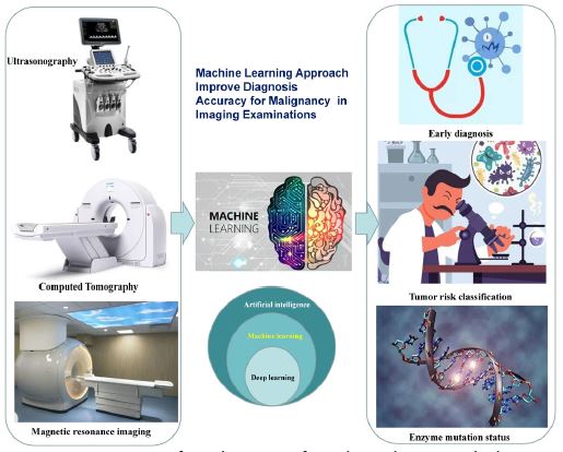
In this mini-review, we provide an overview of the diagnosis and management of malignant tumor using MI-based imaging examinations. Integration of clinical data with imaging features using an ML-based approve has been applied for personalized and predictive medicine in the field of cancer diagnosis. First, ML-based ultrasound, CT and MRI could evaluated early diagnosis value of small size tumor. Second, ultrasound could distinguish primary tumors from metastatic tumors, CT could also play a role in tumor risk classification, and MRI could also predict the pathological grade and enzyme mutation status of malignancy, which can be used to predict early survival and guide clinical decision-making (Figure 1). Third, in the future, it will be possible to implement a predictive model with an ML method and to provide automated decision-making support for the improvement of the patient’s prognosis and the reduction of erroneous clinical diagnosis in routine clinical practice.
References
- Siegel RL, Miller KD, Fuchs HE, Jemal A. Cancer Statistics, 2021 [published correction appears in CA Cancer J Clin. 2021; 71: 359]. CA Cancer J Clin. 2021; 71: 7-33.
- Sung H, Ferlay J, Siegel RL, et al. Global Cancer Statistics 2020: GLOBOCAN Estimates of Incidence and Mortality Worldwide for 36 Cancers in 185 Countries. CA Cancer J Clin. 2021; 71: 209-249.
- Mazzanti R, Arena U, Tassi R. Hepatocellular carcinoma: Where are we? [J]. World journal of experimental medicine. 2016; 6: 21.
- Hack SP, Spahn J, Chen M, et al. IMbrave 050: A Phase III trial of atezolizumab plus bevacizumab in high-risk hepatocellular carcinoma after curative resection or ablation [J]. Future Oncology. 2020; 16: 975-989.
- Yang JD, Hainaut P, Gores GJ, et al. A global view of hepatocellular carcinoma: Trends, risk, prevention and management [J]. Nature reviews Gastroenterology & hepatology. 2019; 16: 589-604.
- Prince D, Liu K, Xu W, et al. Management of patients with intermediate stage hepatocellular carcinoma[J]. Therapeutic Advances in Medical Oncology. 2020; 12: 1758835920970840.
- Schwarze V, Marschner C, Völckers W, et al. Diagnostic value of contrast-enhanced ultrasound versus computed tomography for hepatocellular carcinoma: A retrospective, single-center evaluation of 234 patients [J]. Journal of International Medical Research. 2020; 48: 0300060520930151.
- Li J, Wang J, Lei L, et al. The diagnostic performance of gadoxetic acid disodium-enhanced magnetic resonance imaging and contrast-enhanced multi-detector computed tomography in detecting hepatocellular carcinoma: A meta-analysis of eight prospective studies [J]. European Radiology. 2019; 29: 6519-6528.
- Min JH, Kim JM, Kim YK, et al. Magnetic resonance imaging with extracellular contrast detects hepatocellular carcinoma with greater accuracy than with gadoxetic acid or computed tomography [J]. Clinical Gastroenterology and Hepatology. 2020; 18: 2091-2100. e7.
- Jiang X, Hui F, Qin X, et al. Diagnosis Accuracy and Prognostic Significance of the Dickkopf-1 Protein in Gastrointestinal Carcinomas: Systematic Review and Network Meta-analysis. J Cancer. 2020; 11: 7091-7100
- Zhang Y, Gao J, Bao Y, et al. Diagnostic accuracy and prognostic significance of osteopontin in liver cirrhosis and hepatocellular carcinoma: a Meta-analysis. Biomarkers. 2022; 27: 13-21.
- Abdel Razek AAK, Alksas A, Shehata M, et al. Clinical applications of artificial intelligence and radiomics in neuro-oncology imaging [J]. Insights into Imaging, 2021; 12: 1-17.
- Ahn JC, Qureshi TA, Singal AG, et al. Deep learning in hepatocellular carcinoma: Current status and future perspectives [J]. World Journal of Hepatology. 2021; 13: 2039.
- Vobugari N, Raja V, Sethi U, et al. Advancements in Oncology with Artificial Intelligence—A Review Article [J]. Cancers. 2022; 14: 1349.
- Lawal AI, Kwon S. Application of artificial intelligence to rock mechanics: An overview [J]. Journal of Rock Mechanics and Geo technical Engineering. 2021; 13: 248-266.
- Yazhini K, Loganathan D. A state of art approaches on deep learning models in healthcare: An application perspective [C]//2019 3rd International Conference on Trends in Electronics and Informatics (ICOEI). IEEE, 2019: 195-200.
- Nasir V, Sassani F. A review on deep learning in machining and tool monitoring: methods, opportunities, and challenges [J]. The International Journal of Advanced Manufacturing Technology. 2021; 115: 2683-2709.
- Miotto R, Wang F, Wang S, et al. Deep learning for healthcare: Review, opportunities and challenges [J]. Briefings in bioinformatics. 2018; 19: 1236-1246.
- Akkus Z, Cai J, Boonrod A, Zeinoddini A, Weston AD, et al. A Survey of Deep-Learning Applications in Ultrasound: Artificial Intelligence-Powered Ultrasound for Improving Clinical Workflow. J Am Coll Radiol. 2019; 16: 1318-1328.
- Wu T, Sultan LR, Tian J, Cary TW, Sehgal CM, et al. Machine learning for diagnostic ultrasound of triple-negative breast cancer. Breast Cancer Res Treat. 2019; 173: 365-373.
- Park YR, Kim YJ, Ju W, Nam K, Kim S, Kim KG, et al. Comparison of machine and deep learning for the classification of cervical cancer based on cervicography images. Sci Rep. 2021; 11: 16143.
- Mao B, Ma J, Duan S, Xia Y, Tao Y, et al. Preoperative classification of primary and metastatic liver cancer via machine learning-based ultrasound radiomics. Eur Radiol. 2021; 31: 4576-4586.
- Yu KH, Lee TM, Yen MH, Kou SC, Rosen B, et al. Reproducible Machine Learning Methods for Lung Cancer Detection Using Computed Tomography Images: Algorithm Development and Validation. J Med Internet Res. 2020; 22: e16709.
- Park EK, Lee KS, Seo BK, Cho KR, Woo OH, et al. Machine Learning Approaches to Radiogenomics of Breast Cancer using Low-Dose Perfusion Computed Tomography: Predicting Prognostic Biomarkers and Molecular Subtypes. Sci Rep. 2019; 9: 17847.
- Yin RH, Yang YC, Tang XQ, Shi HF, Duan SF, Pan CJ. Enhanced computed tomography radiomics-based machine-learning methods for predicting the Fuhrman grades of renal clear cell carcinoma. J Xray Sci Technol. 2021; 29: 1149-1160.
- Hu J, Zhao Y, Li M, Liu Y, Wang F, et al. Machine-learning-based computed tomography radiomic analysis for histologic subtype classification of thymic epithelial tumours. Eur J Radiol. 2020; 126: 108929.
- Yu Y, He Z, Ouyang J, Tan Y, Chen Y, et al. Magnetic resonance imaging radiomics predicts preoperative axillary lymph node metastasis to support surgical decisions and is associated with tumor microenvironment in invasive breast cancer: A machine learning, multicenter study. EBio Medicine. 2021; 69: 103460.
- Tahmassebi A, Wengert GJ, Helbich TH, Bago-Horvath Z, Alaei S, et al. Impact of Machine Learning With Multiparametric Magnetic Resonance Imaging of the Breast for Early Prediction of Response to Neoadjuvant Chemotherapy and Survival Outcomes in Breast Cancer Patients. Invest Radiol. 2019; 54: 110-117.
- Hectors SJ, Chen C, Chen J, Wang J, Gordon S, et al. Magnetic Resonance Imaging Radiomics-Based Machine Learning Prediction of Clinically Significant Prostate Cancer in Equivocal PI-RADS 3 Lesions. J Magn Reson Imaging. 2021; 54: 1466-1473.
- Kandemirli SG, Kocak B, Naganawa S, Ozturk K, Yip SSF, et al. Machine Learning-Based Multiparametric Magnetic Resonance Imaging Radiomics for Prediction of H3K27M Mutation in Midline Gliomas. World Neurosurg. 2021; 151: e78-e85.