
Journal of Clinical Images and Medical Case Reports
ISSN 2766-7820
Short Report - Open Access, Volume 3
Once in a blue moon: Unexpected
complication of phaco surgery
Hajar Hnich*; Sarah Aguenaou; Louai Serghini; Zakia Hajji; Amina Berraho
Ophtalmology B Department, Hospital of Specialties, Rabat, Morocco.
*Corresponding Author : Hnich Hajar
Medical Doctor and Resident in Ophthalmology B Department, Hospital of Specialties, 51000, Rabat Morocco.
Tel: +21-263-601-9034;
Email: hajar.hnich93@gmail.com
Received : Oct 08, 2022
Accepted : Oct 28, 2022
Published : Nov 04, 2022
Archived : www.jcimcr.org
Copyright : © Hajar H (2022).
Keywords: Phaco-emulsification complication; Intra ocular lens opacification.
Citation: Hnich H, Aguenaou S, Serghini L, Hajji H, Berraho A, et al. Once in a blue moon: Unexpected complication of phaco surgery. J Clin Images Med Case Rep. 2022; 3(11): 2139.
Introduction
Each year, about 6 million cataract surgeries with Intraocular Lens (IOL) implantation are accomplished [1]; IOL implantation into the human eye during this surgery has been an enormously successful procedure [2].
IOL opacification is a rare condition, it could happen intraoperatively or postoperatively. Up to now, IOL opacifications are described in different IOL materials and designs such as silicone, poly-methyl-methacrylate, hydrophilic acrylic, hydrophobic acrylic and hydrophilic acrylic with hydrophobic coating lenses [1].
Different causes such as the patient’s related conditions, the manufacturing course, and the process of IOL storage, the surgical technique, or sometimes a combination of these factors may lead to clinically significant opacification [3].
We report an exceptionnal peroperative opacification of IOL through the description of a clinical case.
Case report
A 64-year-old male patient with a history of a well controlled type 2 diabetes discovered 10 years ago, presented to our department with decreased visual acuity of both eyes especially the left eye since several months before.
Ophthalmologic examination found the best corrected visual acuity reduced to 4/10 in the right eye, and motion of fingers in the left eye, the slit lamp examination found no abnormalities in the anterior segment except for a dense white cataract preventing the examination of the posterior segment of the left eye and a nuclear sclerosis cataract of the right eye, The dilated fundoscopy was normal in this eye especially no signs of diabetic retinopathy were noticed; the intraocular pressure was within normal in both eyes.
Ocular mode B echography was done in the left eye showing no abnormalities.
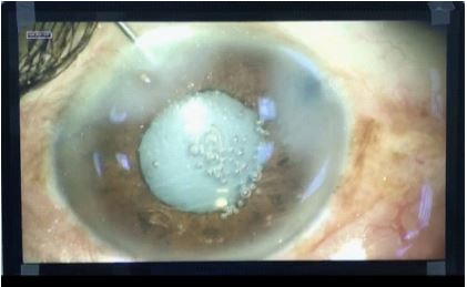
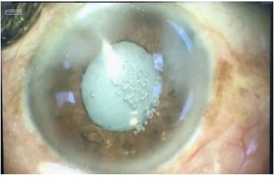
Uneventful cataract surgery was performed on the left eye using a stop and chop technique. a +21 Dioptry foldeble hydrophilic acrylic Intraocular Lens (IOL, manifactured by: CARE GROUP SIGHT SOLUTION PRIVATE LIMITED) was implanted in the bag. The visco elastic was completely washed out from the capsular bag and we were just about to seal the incisions, we noticed sudden cloudy IOL (Figures 1,2). However, we decided not to explant the cloudy IOL, the lens was then not changed. Intra camerular cefuroxime was injected and the incision was hydro sutured.
Discussion
The opacification of hydrophilic acrylic IOL is a rare complication usually occurring in the late postoperative period. the exact causes and pathomechanisms leading to opacification is unknown.
Neuhann et al defined three types of IOL opacifications due to precipitates consisting of calcium and phosphate complexes on the IOL surface and subsurface, respectively Calcium Bicarbonate (CaHPO4) and Hydroxyap-atite (Ca5(PO4)3(OH)) are among the possible complexes that form the opacification. Protein precipitates seem to play a minor role (type 3, ‘pseudocalcification’). Primary IOL opacification—herein referred to as type 1—are supposed to be caused by the IOL itself and usually occur in distinct IOL types or distinct production series. Secondary IOL opacification is herein referred to as type 2 [4].
In our case, under topical anesthesia, the phaco emulsification surgery was uneventful, we used viscoelastic hyaluronate (2%), balanced sel serum and intracameral cefuroxime (1 mg/0.1 ml). We made sure to investigate the cause of opacification and we found that the IOL was kept in a cold storage making the rapide temperature change a probable mechanism of the acute opacification of the lens. other studies have also suggested that acute opacification may occur due to rapid temperature change, and in this situation, there is no need to replace the IOL [3,5].
References
- Werner L. Causes of intraocular lens opacification or discoloration. J Cataract Refract Surg. 2007; 33: 713–726.
- Apple DJ, Ridley Harold. FRCS: A golden anniversary celebration and a golden age. Arch Ophthalmol. 1999; 117: 827–828.
- Sonbolestan SA, Abtahi ZA. Transient intraocular lens opacification during phacoemulsification surgery. J Curr Ophthalmol. 2018; 31: 342-344.
- Mackert M, Muth DR, Vounotrypidis E, et al. Analysis of opacification patterns in Intraocular Lenses (IOL) BMJ Open Ophthalmology. 2021; 6: e000589.
- Helvacı S. Acute opacification of hydrophobic acrylic intraocular lens during implantation: Result of temperature variation. Arq Bras Oftalmol. 2015; 78: 267.