
Journal of Clinical Images and Medical Case Reports
ISSN 2766-7820
Clinical Image - Open Access, Volume 3
Clinical diagnosis of endocarditis
Daniela Casanova*; Ana Sá; Ana Ferreira; Mariana Formigo; Liliana Oliveira; Sara Freitas; Jorge Cotter
Internal Medicine Department, Hospital Senhora da Oliveira, Guimarães, Portugal.
*Corresponding Author : Daniela Casanova
Internal Medicine Department, Hospital Senhora da Oliveira, Cutileiros Street 114, 4835-044, Guimarães, Portugal.
Email: daniela.casanova04@gmail.com
ORCID ID: 0000-0003-2008-9025
Received : Oct 12, 2022
Accepted : Oct 28, 2022
Published : Nov 04, 2022
Archived : www.jcimcr.org
Copyright : © Casanova D (2022).
Abstract
Endocarditis is an entity witch severity and outcome are time related. Sometimes its presentation is linear, such as the one we present on this image, and the echocardiogram is not needed to do the diagnosis. However sometimes this isn’t possible. With this image we pretend to show some typical findings of objective exam and CT scans, so if this appears on emergency department, endocarditis diagnosis is assumed, and prompt treatment is started in order to achieve a good outcome.
Keywords: Endocarditis; Osler nodes; Janeway lesions; Infectious diseases.
Abbreviations: CT: Computed Tomography;
Citation: Casanova D, Sá A, Ferreira A, Formigo M, Oliveira L, et al. Clinical diagnosis of endocarditis. J Clin Images Med Case Rep. 2022; 3(11): 2142.
Case presentation
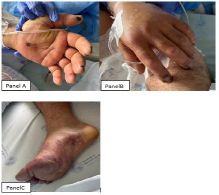
A 57-year-old man with a history of subarachnoid cyst presented to the emergency department with altered mental status and fever with an evolution of 3 days. On physical examination, dark lesions on the palm of both hands and fingertips was noted. On blood analysis there was a thrombocytopenia of 20000/ uL, elevated inflammatory parameters and it was isolated a Staphylococcus aureus on blood cultures. It was performed a cerebral CT that showed bilateral ischemic lesions and a frontal intraparenchymal hematoma, and abdominopelvic CT that showed splenic enfarction. What is the most likely diagnosis?
Discussion/conclusion
The images that we present show typical lesions present on endocarditis, like Osler nodes and Janeway lesions (Panel A and B). And shows an ischemic event decurrent of systemic embolization, on the image presented by foot ischemia (Panel C), and on computed tomography with splenic infarction. These clinical signs made the clinical diagnosis of endocarditis, even without doing an echocardiogram.