
Journal of Clinical Images and Medical Case Reports
ISSN 2766-7820
Case Report - Open Access, Volume 3
A masquerading ovarian mass
MD Ray*; Manish Kumar Gaur
Department of Surgical Oncology, DR BRA-IRCH, All India Institute of Medical Sciences, New Delhi, India.
*Corresponding Author : MD Ray
Department of Surgical Oncology, DR BRA-IRCH, All India Institute of Medical Sciences, New Delhi, India.
Email: Dr_mdray@yahoo.com
Received : Oct 12, 2022
Accepted : Oct 31, 2022
Published : Nov 07, 2022
Archived : www.jcimcr.org
Copyright : © MD Ray (2022).
Citation: Ray MD, Gaur MK. A masquerading ovarian mass. J Clin Images Med Case Rep. 2022; 3(11): 2144.
Description
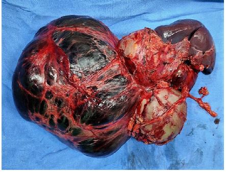
A 23 years old lady presented with lower abdominal vague pain and fullness for 6 months. Physical examination revealed a lump in the left lumbar region extending into the pelvis. The upper border was palpable, but the lower limit was not palpable. Computerized tomography of the abdomen revealed a multiloculated cystic lesion with minimal enhancement probably arising from the left adnexa. The right adnexa, uterus, pancreas, spleen appeared normal. Serum tumor markers: CA- 125, alpha-fetoprotein, beta HCG, LDH, and CA-19.9 were within normal limits. Diagnosis of left adnexal complex cystic was made and planned for Fertility preserving staging laparotomy. Intraoperatively, bilateral adnexa and uterus were normal. A large soft multicystic lesion measuring 15 x 10 x 7 cm was noted arising from the tail of the pancreas (Figure 1). No other lesions were noted in the peritoneal cavity. Distal pancreatectomy with splenectomy was performed to achieve R-0 resection. The post-operative hospital stay was uneventful, and the patient was discharged on POD-5. The final histopathological report revealed a multiloculated cyst showing irregular lymphovascular spaces lined by flattened, bland cells within fibroblastic and collagenous stroma suggestive of consistent with cystic lymphangioma of the pancreas. On follow-up, the patient is doing well after 1 year of the surgical excision.
Cystic lymphangiomas of the pancreas are very rare, often asymptomatic, and have a female preponderance. The preoperative diagnosis is seldom made and is often diagnosed after the surgery. The imaging findings occasionally reveal a multilocular well-defined cystic lesion with a homogenous composition which shows enhancement post-contrast injection and is well appreciated on CT and MRI. The common differential diagnosis includes simple cysts, pseudocysts, serous cysts, Intraductal Mucinous Neoplasm (IPMN), and Mucinous Cystic Neoplasm (MCN). Although considered benign neoplasms, a few of them may be locally invasive; thus, the treatment of choice for these lesions remains complete surgical excision. Following a complete excision, the prognosis is excellent, with a very low incidence of recurrence.