
Journal of Clinical Images and Medical Case Reports
ISSN 2766-7820
Case Report - Open Access, Volume 3
A rare case of intramedullary osteosclerosis and literature review
Saoussen Miladi; Yosr El Mabrouk*; Alia Fazaa; Hiba Boussaa; Kawther Ben Abdelghani; Ahmed Laatar
Rheumatology Department, Faculty of Medicine of Tunis, Mongi Slim Hospital, University of Tunis El Manar, Tunisia.
*Corresponding Author : Yosr El mabrouk
Rheumatology Department, Faculty of Medicine of Tunis, Mongi Slim Hospital, University of Tunis El Manar, Tunisia.
Email: yosr.elmabrouk@gmail.com
Received : Oct 13, 2022
Accepted : Nov 03, 2022
Published : Nov 10, 2022
Archived : www.jcimcr.org
Copyright : © Mabrouk Y (2022).
Abstract
Intramedullary Osteosclerosis (IMOS) is a rare idiopathic dysplasia of the long bones shafts. IMOS remain an un known diagnosis because of its rarity and the absence of specific features. Indeed, the diagnosis of IMOS can be confirmed only after excluding other differential diagnosis of osteosclerotic lesions. We report here a case of IMOS of the tibia shafts, diagnosed in a 53-year-old man. Medical history did not reveal a history of trauma or infection or familial bone disease and all other causes of osteosclerosis were excluded. The X-Rays showed extensive sclerotic intramedullary lesion of the tibia shafts, cortical bone thickening, and narrowing of the medullary cavity. No periosteal reaction or soft-tissue abnormality or fracture line were noted. The radiological signs were not pathognomonic, but the clinic, biology, histology, as well as the absence of medical or family history strongly suggested the diagnostic.
Citation: Milad S, Mabrouk YE, Fazaa A, Boussaa H, Abdelghani KB, et al. A rare case of intramedullary osteosclerosis and literature review. J Clin Images Med Case Rep. 2022; 3(11): 2151.
Introduction
Intramedullary Osteosclerosis (IMOS) is a sclerosing dysplasia associated with increased bone formation in the medullary cavity. The term intramedullary osteosclerosis was introduced by Abdul-Karim et al in 1988 to describe a rare pathology associated with new bone formation localized mainly in the diaphysis of the tibia in adult female patients [1]. It is a rare disease, without specific radiological signs except for the osteosclerotic lesion and it is not associated with a family history, infection, trauma or systemic disease [2]. Although the diagnosis of IMOS is confirmed after excluding other sclerotic lesions, it is not well recognized due to its rarity and absence of specific signs. Consequently, these situations can lead to a delay in the diagnosis. Here, we report the clinical and radiological characteristics of a patient diagnosed with IMOS.
Case presentation
A 53-year-old man with a history of diabetes and hypertension, was followed since 2020 for a severe spontaneous pain on the left leg without triggering factor, mild at first, then getting worse gradually. Pain was prolonged, chronic and recurrentand was worsened by physical activity. He did not report any illness of the right leg. Nonsteroidal Anti-Inflammatory Drugs (NSAID) were ineffective. The patient did not report any notable trauma or infection or familial bone disease history.
At examination, we did not notice local heat, swelling, or redness. Laboratory data were within normal limits: erythrocyte sedimentation rate (ESR): 20 mm, C-Reactive Protein (CRP): 5 mg/l, normal liver and kidney function, calcemia: 2.3 mmol/l, phosphoremia: 1.18 mmo/l, alkaline phosphatase: 58 UI/L.
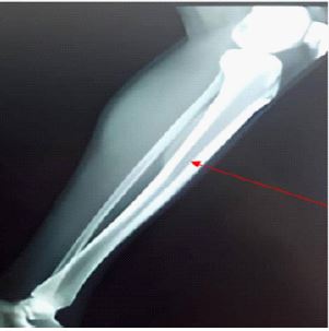
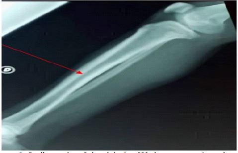
Plain Radiographies showed bilateral massive sclerotic intramedullary lesion of tibial shafts, cortical bone thickening, and narrowing of the medullary cavity. No periosteal reaction or soft-tissue abnormality or fracture lines were seen (Figures 1&2). The diagnosis of IMOS of both tibia shafts was suspected. As etiological diagnosis of the osteosclerosis, we discussed malignant tumors (osteosarcoma, lymphoma, bone metastasis), benign tumors such as multifocal osteoid osteoma, stress fractures, metabolic disorders and hereditary bone diseases. Nevertheless, all these diagnosis were excluded aslaboratory findings were normal, no familial bone disease history was present, no extensive periosteal new bone formation and soft-tissue abnormality were noted on the radiography. Indeed, the bilateral characteristics of the process, and the exclusion of other diagnosis were sufficient to retain the diagnosis of IMOS and we did not perform any supplementary exploration. Concerning therapeutic management, the patient received level 2 analgesic and NSAID with a good response.The last out patient meeting was held on February 2022 and he reported a good evolution.
Discussion
We report here a case of IMOS in a 53-year-old man. To the best of our knowledge, there are less than twenty cases reported in literature.
In the series of Kullanuch Chanchairujira et al, nine female patients with leg pain and imaging signs of IMOS were detected during a 25-years period [3]. Kensaku Abe et al reported three cases (one man, two women) of osteosclerotic lesions (femoral diaphyses, tibial diaphysis) examined between 2015 and 2019 [2]. Vasilios Skiadas et al presented the case of a 60-year-old woman with a bilateral midfemoral pain for 8 years [4]. In the case of Balkissoon et al, a 39-year-old woman experienced intermittent severe pain in both lower legs for several years [5].
These data suggest that this condition is more common in women than in men unlike our case.
No similar musculoskeletal illness was found in any patient’s family history. None of these patients have a history of trauma or infection. No abnormal laboratory results including inflammatory reaction were observed [2-4], such in our case.
The diagnosis of intramedullary osteosclerosis was retained after the exclusion of disorders related to osteosclerosis, including osteosarcoma, lymphoma, metastasis, stress fractures, metabolic disorders and other sclerosing bone dysplasia [1,5]. Metabolic disorders such as renal osteodystrophy, hypervitaminosis A and pseudohypoparathyroidism need to be considered. Generalized osteosclerosis is more characteristic of renal osteodystrophy and pseudohypoparathyroidism. Hypervitaminosis A may lead to a periosteal bone formation. Normal laboratory findings and clinical features can eliminate these causes [3,9]. Only two known conditions, Camurati-Engelmann disease and Ribbing disease are presented with radiological and scintigraphic peculiarities similar to those of osteosclerosis intramedullary. But, they can easily be dismissed since the two are hereditary: (Engelmann: autosomal dominant, Ribbing: autosomal recessive) [10,11].
Radiographs revealed, in the case of Vasilios et al, diffuse bilateral asymmetric diaphyseal sclerosis of femoral bones, predominantly endosteal, more extensive on the left side, with mild expansion and cortical thickening of the affected segments. No periosteal reaction was seen [4]. In the case of Balkissoon et al, The radiographs demonstrated bilateral asymmetric mid-tibial and minimal fibular shaft endosteal cortical scleroses. There was no substantial soft-tissue abnormality [5]. In the cohort of Kullanuch Chanchairujira et al, 16 bone lesions were identified. All were located in lower extremities. Both lower extremities were involved in the majority of patients. A single extremity was involved in two patients. Cortical thickening was seen in 10 bones and soft-tissue swelling was seen in eight lesions. No periosteal reaction was seen [3]. Radiographs of the legs showed bilateral hyperostosis involving the mid-third of the tibias in the case of Rosenberg et al [6] and diffuse diaphyseal sclerosis of the femur with widening of bone and endosteal hyperostosis in the case of Puranik et al [7]. The clinical and radiographic findings in our patient supported those studies.
Computed Tomography (CT) scans showed, in these series intramedullary sclerosis and soft-tissue swelling adjacent to the cortex. The cortex was intact but thickened [2-4,8]. In the case of Balkissoon et al, CT scan proofs were not available such as in our case [5].
Concerning MRI findings, MR images showed low signal intensity on images obtained with all pulse sequences, minimal increased signal intensity was seen on T2-weighted images. Gadolinium-enhanced T1-weighted fat-saturated images were obtained, and the images revealed minimal enhancement in the lesions or surrounding soft tissue [2-4]. In our case, MRI findings were not available.
Skeletal scintigraphy was performed in five patients with nine lesions in the series of Chanchairujira et al. It showed intense tracer uptake corresponding to the abnormalities seen on conventional radiographs [3,4,6]. In one patient with bilateral lesions, an asymptomatic lesion was incidentally found in an area that appeared normal at conventional radiography [3]. Abe K et al and Puranik et al found that whole body 99 mTc-Methylene Diphosphonate (MDP) bone scan showed abnormal tracer uptake with intense sclerosis in the intramedullary region. Moreover, triphasicbone scan was performed: the initial vascular phase and blood pool images at 2 min (phase 2) showed no evidence of increased vascularity or soft tissue tracer pooling. Delayed bone images (phase 3) showed a fusiform-shaped intense area of tracer uptake in the diaphysis [2,7]. In our setting, scintigraphy was not performed.
Open biopsy was carried out and histologic findings were available in the series of Chanchairujira et al, but were non-specific. The peripheral and major part of the lesion was characterized by the replacement of normal spongiosa by markedly sclerotic trabeculae and were seen radiographically as osteosclerosis. There were some areas of osteoblastic rimming of the trabeculae. Centrally, cell proliferation associated with immature collagen deposition was noted [3]. In the cases of Abe K et al, the histology showed trabecular bone sclerosis with hypocellular fibrous tissue. The culture was negative [2]. Culture was sterile in the case of Vasilios et al. Pathologic examination revealed a replacement of normal spongiosa by markedly sclerotic and thickened trabeculae, obviously, seen radiographically as osteosclerosis, in the case of Puranik too [4,7].
The management of intramedullary osteosclerosis is not well described in the literature and is not yet standardized.
NSAID seem to be effective in this disease. Indeed, in our case, the patient improved after prescription of diclofenac and he was relieved in the follow up. In the case of Skiadas et al, the patient also showed improvement following NSAID therapy administered for one month [4]. In the series of Abe et al, pain was uncontrolled by even opioid use [2]. In our case, the association of NSAID and level 2 analgesic was sufficient.
Open surgical biopsy seems to be also an efficient treatment of IMOS. In fact, besides the diagnosis confirmation, this technique allowed medullary decompression. In the Kensaku et al series, open biopsy was able to reduce the intensity pain, which is controlled afterwards only by NSAID [2].
Although, there were limitations to our study, such as the lack of CT, MRI, scintigraphy and histologic results, we believe that our study and our review of the literature can add data to this still unknown disease.
Conclusion
As seen above, IMOS should be considered in the differential diagnosis in any unexplained intense pain symptom in the diaphyseal region of long bones. Massive sclerotic lesions seen in X-Rays as well as the absence of medical or family history strongly suggested our diagnostic. NSAID was considered as a relievable treatment.
Declarations
Acknowledgements: None.
Conflicts of interest: None.
Author contribution: Yosr el mabrouk has drafted the work. Saousenmiladi has substantively revised the work. Kaouthar ben abdelghani, Alia Fazaa and hibaboussaa have made substantial contributions to the literature research. Ahmed Laatar has made substantial contributions to the conception of the work.
Ethical approval: None.
Consent: The patient gave her consent for publishing the case with absolute respect of anonymity.
Consent: Written informed consent was obtained from the patient to publish this report in accordance with the journal’s patient consent policy.
References
- Abdul Karim FW, Carter JR, Markley JT, et al. Intramedullary osteosclerosis: Report of the clinicopathologic features of five cases. Orthopedics. 1988; 11: 1667–1675.
- Abe K, Yamamoto N, Hayashi K, Takeuchi A, Miwa S, et al. Diagnosis and treatment of intramedullary osteosclerosis: A report of three cases and literature review. BMC Musculoskeletal Disorders. 19; 21: 762.
- Chanchairujira K, Chung CB, Lai YM, Haghighi P, Resnick D, et al. Intramedullary osteosclerosis: imaging features in nine patients. Radiology. 2001; 220: 225-230.
- Vasilios Skiadas, Minos Tyllianakis, Vasiliki Zolota, Apostolos Karantanas. Intramedullary osteosclerosis: A case report and literature review. The American Journal of Orthopedics. 2012; 41: 496-499.
- Balkissoon ARA, Hayes CW. Case 14: Intramedullary Osteosclerosis. Radiology. 1999; 212: 708–710.
- Rosenberg C, Laredo JD, Rozenberg S, Marre JP, BourgeoisP. Bilateral diaphyseal tibial hyperostosis: A confusing hyperostosis. Joint Bone Spine. 2008; 75: 751–752.
- Puranik, Ameya D et al. Intramedullary osteosclerosis of right femur confirmed on triphasic bone SPECT/CT in a patient with equivocal radiological features. Indian journal of nuclear medicine: IJNM: The official journal of the Society of Nuclear Medicine. India. 2016; 31: 39-41.
- Casagranda B, Heller MT, Costello J. Intramedullary osteosclerosis: An incidental sclerotic lesion in a trauma patient. Radiol Case Rep. 2015; 8: 878.
- Harold G Jacobson, et al. Dense Bone- Too Much Bone: Radiological Considerations and Differential Diagnosis. Skeletal radiol. 1985; 13: 97-113.
- Kaftori JK, Kleinhaus U, Naveh Y. Progressive diaphyseal dysplasia (Camurati-Engelmann): radiographic follow-up and CT findings. Radiology. 1987; 164: 777–782.
- Ribbing S Hereditary, Multiple diaphyseal sclerosis. Acta Radiol. 1949; 31: 522–536.