
Journal of Clinical Images and Medical Case Reports
ISSN 2766-7820
Clinical Image - Open Access, Volume 3
Three layer test in bronchiectasis
KC Shikha; Harsha Sreekumar; Shravan Kumar; Arjun Padmanabhan*
Department of Respiratory Medicine, KIMS Health, Anayara P.O, Trivandrum –695029, Kerala, India.
*Corresponding Author : Arjun Padmanabhan
Senior Consultant, Professor and Head, Department of Respiratory Medicine, KIMS Health, Anayara P.O, Trivandrum –695029, Kerala, India.
Ph: 91-471-3041322, 91-9447151919,
Fax: 91-471-2446535;
Email: dr.p.arjun@gmail.com
Received : Oct 19, 2022
Accepted : Nov 04, 2022
Published : Nov 11, 2022
Archived : www.jcimcr.org
Copyright : © Padmanabhan A (2022).
Citation: Shikha KC, Sreekumar H, Kumar S, Padmanabhan A. Three layer test in bronchiectasis. J Clin Images Med Case Rep. 2022; 3(11): 2153.
Description
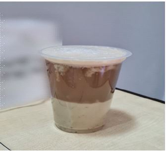
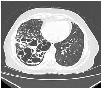
A 52 year old woman having bronchiectasis, presented to us with worsening cough which was productive with profuse, foul smelling sputum of a week’s duration. A clinical diagnosis of infective exacerbation of bronchiectasis was made. She was started on antibiotics based on her previous sputum culture sensitivity reports. She was asked to collect an entire day’s sputum in a transparent container which demonstrated the classical three layer sputum (Figure 1). Her high resolution CT scan of the thorax was suggestive of cystic bronchiectasis, predominantly involving the right lung (Figure 2). Some of the cysts contained fluid levels also, indicating active infection.
The three layer sputum consists of a foamy upper layer, mucous middle layer, and viscous purulent bottom layer [1]. The bottom layer consists of cell debris and sometimes Dittrich plugs may also be seen [2]. These are greyish white foul smelling masses of bacterial and fatty acid crystals, typically seen in suppurative lung disease. Her sputum grew Pseudomonas aeuaeuroginosa and she responded promptly to appropriate antibiotic therapy. The three layer sputum is seldom seen in the modern era with the advent of potent antibiotics. In olden days, it used to be done as a bedside test for diagnosis of suppurative lung diseases, especially bronchiectasis.
Declarations
Funding statement: This research has not received any specific grant from any funding agency in the public, commercial or not-for-profit sectors.
Competing interests statement: We, the authors declare that there are no competing interests present as far as this submission for publication is concerned.
Contributorship statement:Skihka KC, Harsha Sreekumar, Shravan Kumar and Arjun Padmanabhan were initially involved in workup of the patient, treatment and further management.
References
- Rademacher J, Welte T: Bronchiectasis – diagnosis and treatment. Dtsch Arztebl Int. 2011; 108: 809–815.
- Rafael Martinez-Giron, Santiago Martinez-Torre. Dittrich’s plugs in sputum: Morphological observations. Cytopathology. 2012; 23: 278-279.